The immense water-absorbing capacity of hydrogels has made them exciting materials in a number of fields including engineering and biotechnology. It is possible to design ‘smart’ hydrogels, where their water-binding properties can be tuned by changing the environmental conditions of the hydrogel, such as by applying a voltage.

Image Credit: Gilmanshin/Shutterstock.com
Hydrogels are materials that can absorb and retain large amounts of water. To achieve this, hydrogels have a three-dimensional network structure that is strongly hydrophilic and creates a large, attractive surface area for binding water. Hydrogel materials can be crystalline, amorphous, or semicrystalline, and the underlying polymer structure is crucial for the performance of the hydrogel material.
Probably the most widely used application of hydrogels is in contact lens making, where the high water content of the material helps avoid drying the surface of the eye during wear.
Other medical applications for hydrogels include wound dressing and drug delivery. For wound dressings, hydrogel polymers are permeable to oxygen and can help absorb fluids and cells that are exuded from the wound.
They can also provide an antiseptic effect to prevent infection and help maintain a fluid balance at the wound site to promote healing.
Optical Spectroscopy of Hydrogels
As the structure of the polymer is key to the performance of the hydrogel, analytical techniques that can evaluate both the chemical composition and arrangement of the polymer chains is necessary for the analysis and development of new hydrogels.
Ideally, the porous network structure of the hydrogel would be tailored for the application to make sure it can remove the correct waste and provide the optimal moisture levels.
Doing this means needing to use advanced characterization techniques.
Raman spectroscopy has been one of the workhorse techniques of pharmaceutical and chemical manufacturing. It is an excellent quantitative and qualitative technique that can recover information on both the chemical identity and bonding structure of molecules in solution as well as solids.
One advantage of Raman spectroscopy is that it is a label-free technique so requires minimal additional sample preparation for measurements.
It can also measure the system in its unperturbed state, without any potential distortion from interactions and binding with the labeling species.
Hydrogels are typical in heavily aqueous environments, whereas the strong solvent response in the infrared can make infrared absorption spectroscopy challenging. Raman spectroscopy typically suffers from less spectral interference from the solvent.
However, Raman spectroscopy on hydrogels suffers from several drawbacks that have limited its uptake for these materials to date. The main one of these is the large fluorescence signal that is generated by samples following photoexcitation with a laser.
As the Raman effect is relatively weak, the large fluorescence background can swamp the smaller Raman signals.
Unfortunately, there is also a challenging compromise to be made in terms of the choice of the incident laser wavelength.

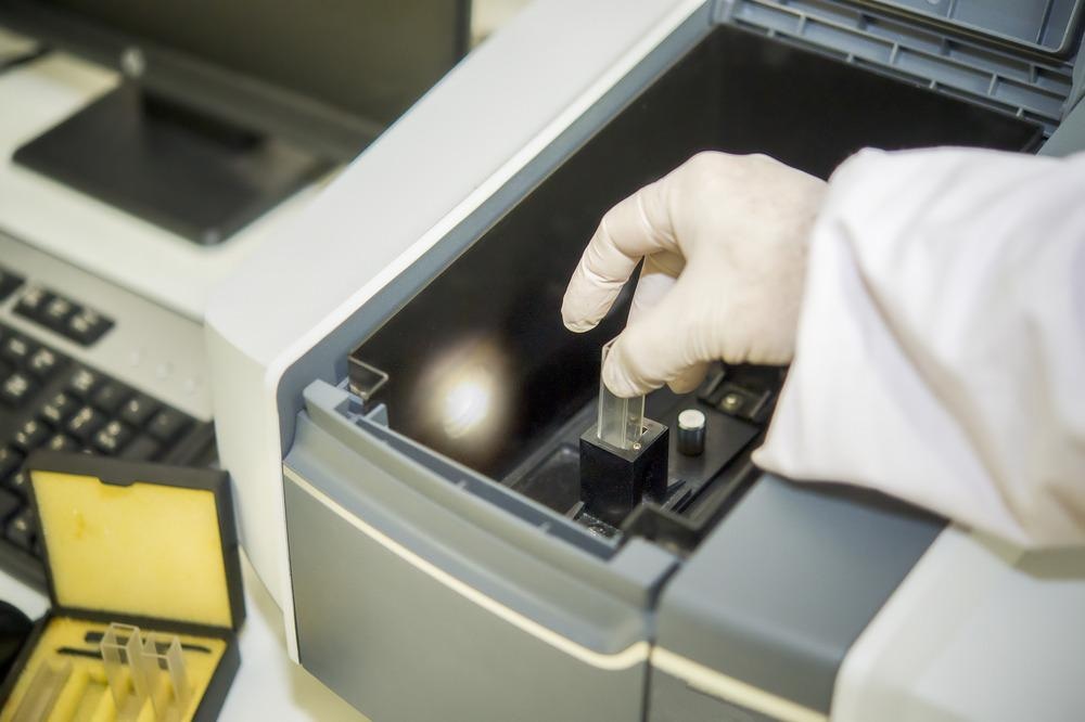
Image Credit: Rabbitmindphoto/Shutterstock.com
While shorter wavelengths give a stronger Raman signal, they often create an even greater fluorescence background and result in poorer contrast.
Upgrading Raman Spectroscopy
One approach to circumvent the challenges associated with traditional, steady-state Raman spectroscopy is to perform an optical Kerr gate measurement instead.
Kerr gate Raman measurements take advantage of the lifetime differences between the fluorescence and Raman signals.
Optical Kerr gate Raman spectroscopy works by using a picosecond, or shorter excitation pulse.
Rather than recording continuously, the detectors are set to only record signals during the laser pulse itself.
This is an excellent way to suppress any background signals from fluorescence or ambient light, but requires electronic communication speeds fast enough that detectors can be triggered and record over relatively short (< 1 ns) timescales.
While optical Kerr gate Raman has been used in laboratory settings, the complexity of the equipment and the relatively bulky size of set-ups has limited its use outside of specialist groups.
However, a recent development in using time-gated single-photon avalanche photodiodes (SPADs) may bring the technique to a wider audience.4
Kerr Gate Raman on Hydrogels
The research team were able to use a time-resolved SPAD sensor to investigate the diffusion properties of two species (Metronidazole and Vitamin C) in a specific hydrogel and as a function of varying substrate concentrations.
By obtaining both background-free Raman spectra and fluorescence lifetimes, the researchers could investigate whether drugs were binding at certain points in the hydrogel or undergoing some type of degradation.
All of this information could be used to map the diffusion paths of the substrates in the hydrogel and recover information on likely interactions and drug-release timescales.
The aqueous nature of hydrogels makes them an appealing drug delivery system, so new tools to understand the mechanisms of drug release in their structures are important for further developments in this area.
Optical Kerr gate Raman spectroscopy is an incredibly general technique. This is because it is label-free, and Raman has excellent sensitivity to the vibrational structure of different molecules.
Most molecules have a characteristic ‘fingerprint’ spectrum, so can be adapted for other hydrogel/substrate combinations and, therefore, have great potential as a tool for mechanistic studies in the pharmaceutical industry.
As detector electronics and communication speeds continue to improve and systems are simplified further, Kerr gate Raman may become a more common way of hydrogel monitoring.
References and Further Reading
Ahmed, E. M. (2015). Hydrogel: Preparation, characterization, and applications: A review. Journal of Advanced Research, 6(2), 105–121. https://doi.org/10.1016/j.jare.2013.07.006
Balakrishnan, B., Mohanty, M., Umashankar, P. R., & Jayakrishnan, A. (2005). Evaluation of an in situ forming hydrogel wound dressing based on oxidized alginate and gelatin. Biomaterials, 26(32), 6335–6342. https://doi.org/10.1016/j.biomaterials.2005.04.012
Zini, J., Kekkonen, J., Kaikkonen, V. A., Laaksonen, T., Keränen, P., Talala, T., Mäkynen, A. J., Yliperttula, M., & Nissinen, I. (2021). Drug diffusivities in nanofibrillar cellulose hydrogel by combined time-resolved Raman and fluorescence spectroscopy. Journal of Controlled Release, 334(February), 367–375. https://doi.org/10.1016/j.jconrel.2021.04.032
Disclaimer: The views expressed here are those of the author expressed in their private capacity and do not necessarily represent the views of AZoM.com Limited T/A AZoNetwork the owner and operator of this website. This disclaimer forms part of the Terms and conditions of use of this website.