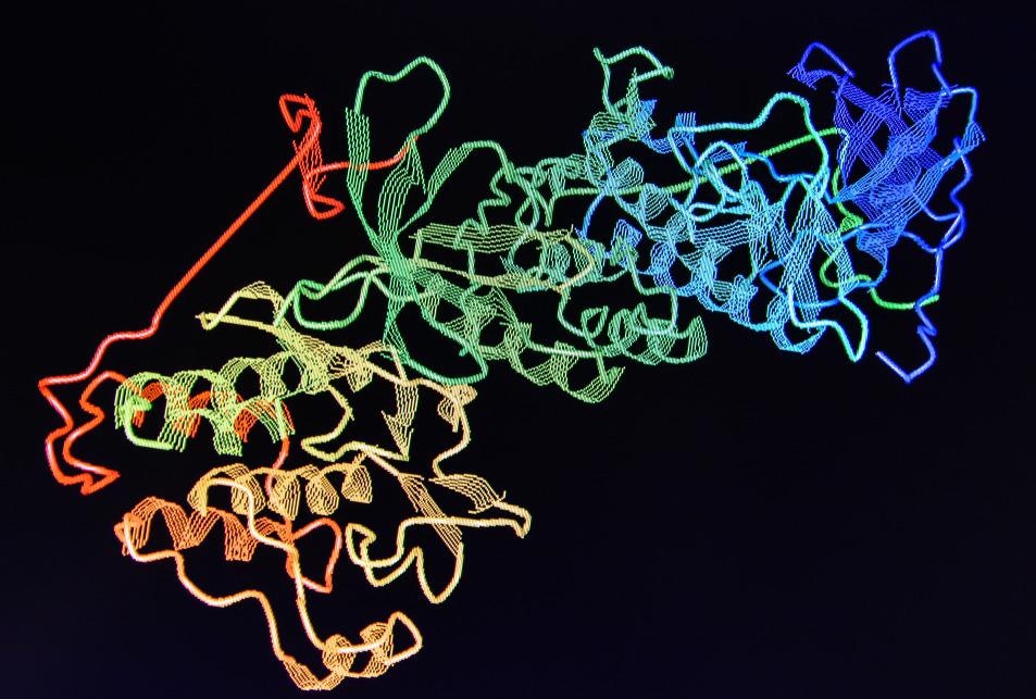
Manipulating the polarization of light in spectroscopies can reveal previously hidden spectral and structural information on the species of interest. One key example of this is the use of circularly polarized light in protein studies to perform circular dichroism measurements.

Image Credit: Sergei Drozd/Shutterstock.com
With over 24,000 papers making use of the power of this technique, circular dichroism has proved an invaluable tool for the structural identification of proteins and recovering information on drug binding and interaction sites.1
The Problem with Chiral Species
Circular dichroism involves looking at how the response of a system differs when exposed to either left or right-handed circularly polarized light.
Many molecules exhibit no circular dichroism signal. However, chiral species will preferentially absorb one state of circularly polarized light resulting in an overall different signal.
A typical example of chiral molecules is amino acids. This chirality is often retained when incorporated into larger protein structures, posing a significant challenge during drug development. Here, different enantiomers of drug molecules do not necessarily interact with the protein in the same way.2
Still, this provides a useful spectroscopic handle to investigate a number of aspects of protein structure.
Current Methods to Research Circular Dichroism
One of the critical applications of circular dichroism for proteins has been in the determination of secondary structure. Different structural arrangements, such as α-helical structures or β-pleated sheets, have characteristic bands in their circular dichroism spectra in the UV region of the electromagnetic spectrum.3
Changes in the band position and spectral shapes in the circular dichroism spectra can be used to monitor protein folding as well as provide estimates on the degree of disorder in the structure. Several databases and documented workflows exist for nearly entirely automated analysis of circular dichroism spectra that can provide predictions of possible structures.1
Circular dichroism has also been one tool of choice in the pharmaceutical industry as a non-destructive technique for quality control on drug and protein production, process optimization, and bio target interaction studies.
Recent Advancements with Complementary Tools
While circular dichroism is a valuable tool in isolation, it is increasingly used alongside complementary theoretical and experimental methods, including molecular dynamics simulations and other structural biology techniques such as X-ray diffraction.1
For example, thermal melt circular dichroism was used alongside the DichroWeb suite of secondary structural analysis tools to perform docking studies to determine the interaction site of valproic acid, an anticonvulsant drug.4
Advancements in theoretical modeling approaches improve the accuracy and speed to determine potential protein structures at a given temperature. Not only that, but these methods enable the development of new tools for the prediction of circular dichroism spectra.
One group of approaches utilize quantum chemical calculations based on crystal structure geometries to simulate the circular dichroism signal5; others, such as the PDB2CD and PDBMD2CD servers, use least-squares fitting to reference datasets for similar proteins.6,7
While the UV region has typically been the spectral range of choice for performing CD measurements, X-ray circular dichroism beamlines at synchrotrons have become increasingly common to allow greater site-specificity in measurements.
Vibrational circular dichroism, which involves measures using infrared light, is another area in active development that has proved especially successful in the study of protein fibrils.8
The Future of Circular Dichroism
UV circular dichroism is already an established workhorse technique in many industries, such as the pharmaceutical industry. The ability of circular dichroism to characterize species like monoclonal antibodies9 means that it is likely to remain a popular tool to support the development and testing of new therapeutics.
As the circular dichroism signal from proteins is typically minimal, developing plasmonic structures to enhance the circular dichroism signature for biostructures may become more common.9 Modeling developments will continue to improve the structural information obtained from measurements. They will likely play a growing role in studies looking to elucidate the mechanisms for drug-substrate interactions.
Challenges to Overcome
Working in the UV region of the electromagnetic spectrum often poses significant challenges for tabletop experiments regarding the optics required. UV circular dichroism has had limited success for species like unsubstituted carbohydrates, where sub-200 nm wavelengths would be necessary for complete structural investigations.
Many solvents have significant absorption in this region, as do buffer systems required to stabilize the proteins and control their environmental conditions, which may cause interference.
Another challenge for circular dichroism measurements is that the measurements are highly sensitive to slight differences in the equipment used and sample preparation.
Guides have been created for best practices, sample preparation, and calibration procedures, but there is still a need for more reproducible measurements across different instruments. While some of the challenges are inherent to the sample types themselves, as proteins can degrade or aggregate readily, others are due to the inherently small signals from proteins. More sensitive instruments for shorter scan times or methods to enhance the protein signal would help address this.
References and Further Reading
Miles, A., Janes, R. and Wallace, B., (2021) Tools and methods for circular dichroism spectroscopy of proteins: a tutorial review. Chemical Society Reviews, 50(15), pp.8400-8413. Available at: https://doi.org/10.1039/d0cs00558d [Accessed August 2021]
H. Brooks, W., C. Guida, W. and G. Daniel, K., (2011) The Significance of Chirality in Drug Design and Development. Current Topics in Medicinal Chemistry, 11(7), pp.760-770. Available at: https://doi.org/10.2174/156802611795165098 [Accessed August 2021]
Greenfield, N., (2006) Using circular dichroism spectra to estimate protein secondary structure. Nature Protocols, 1(6), pp.2876-2890. Available at: https://doi.org/10.1038/nprot.2006.202 [Accessed August 2021]
Zanatta, G., Sula, A., Miles, A., Ng, L., Torella, R., Pryde, D., DeCaen, P. and Wallace, B., (2019) Valproic acid interactions with the NavMs voltage-gated sodium channel. Proceedings of the National Academy of Sciences, 116(52), pp.26549-26554. Available at: https://doi.org/10.1073/pnas.1909696116 [Accessed August 2021]
Drew, E. and Janes, R., (2020) PDBMD2CD: providing predicted protein circular dichroism spectra from multiple molecular dynamics-generated protein structures. Nucleic Acids Research, 48(W1), pp.W17-W24. Available at: https://doi.org/10.1093/NAR/GKAA296 [Accessed August 2021]
Mavridis, L. and Janes, R., (2016) PDB2CD: a web-based application for the generation of circular dichroism spectra from protein atomic coordinates. Bioinformatics, 33(1), pp.56-63. Available at: https://doi.org/10.1093/bioinformatics/btw554 [Accessed August 2021]
Nafie, L., (2020) Vibrational optical activity: From discovery and development to future challenges. Chirality, 32(5), pp.667-692. Available at: https://doi.org/10.1002/chir.23191 [Accessed August 2021]
Moro Pérez, L., Rodríguez Taño, A., Martín Márquez, L., Gómez Pérez, J., Valle Garay, A. and Blanco Santana, R., (2019) Conformational characterization of a novel anti-HER2 candidate antibody. PLOS ONE, 14(5), p.e0215442. Available at: https://doi.org/10.1371/journal.pone.0215442 [Accessed August 2021]
Hou, S., Yan, J., Hu, Z. and Wu, X., (2016) Enhancing the plasmonic circular dichroism by entrapping chiral molecules at the core–shell interface of rod-shaped Au@Ag nanocrystals. Chemical Communications, 52(10), pp.2059-2062. Available at: https://doi.org/10.1039/c5cc08505e [Accessed August 2021]
Disclaimer: The views expressed here are those of the author expressed in their private capacity and do not necessarily represent the views of AZoM.com Limited T/A AZoNetwork the owner and operator of this website. This disclaimer forms part of the Terms and conditions of use of this website.