Image Credit: Benjamin Outram/Shutterstock.com
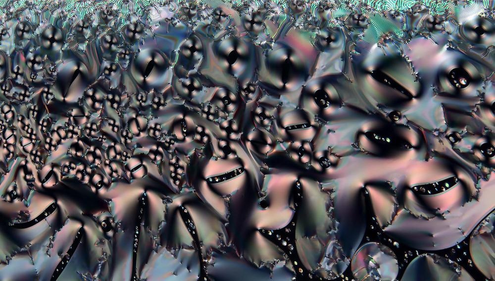
Polarized light microscopy uses polarized light that travels through a double refracting material, then generated into a high-contrast image via the collection of light by a second polarizing filter. The technique has been leveraged by a wide range of industries and adapted for a variety of applications. While it is mainly used in geology, the fields of biology, medicine, industry, and food science have also benefited.
How Does Polarized Microscopy Work?
Polarized light microscopy is considered a contrast-enhancing method that evaluates the three-dimensional structure and composition of anisotropic samples. While conventional light microscopes use unpolarized white light, the type of light that is visible to the human eye and is formed of waves that vibrate in random directions, polarized light microscopy uses polarizing filters to make use of polarized light, which cannot be seen by the human eye and composes waves that vibrate in a single direction.
The use of polarized light in microscopy results in the generation of images of much greater quality than conventional light microscopy when investigating anisotropic materials. Such materials are considered “direction-dependent”, meaning that their behavior depends on the direction. For example, the properties of wood differ depending on whether the action is occurring against or with the wood grain. In comparison, steel, an isotropic material, does not demonstrate this alteration in behavior; it is the same regardless of the direction.
In polarized light microscopy, light waves (omnidirectional) are polarized by a polarizer, forcing the waves to move in just one direction. Therefore, only light waves vibrating in parallel move through, with light waves in all other directions blocked. Light waves are then emitted through the sample and vibrated in an east-west orientation. The waves continue upwards undisturbed until the analyzer is switched on, at which point, only light moving in a north-south position is allowed through. The placement of a second polarizer (or analyzer) in the light’s path at an orientation of 90 degrees, in relation to the axis of the first polarizer, blocks out all light. The polarizers are “crossed’ as all east-west and north-south directions are blocked.
Applications of Polarized Light Microscopy
Polarized light microscopy is used to highlight the characteristic of various anisotropic samples. It is commonly used to investigate crystals, bone, fibers, and minerals, although it can be used to explore a wide range of materials, with new applications emerging each year. The two filters can be adjusted to allow light at different angles (as described above) so that the sample can be visualized from different perspectives.
Biological applications
Double-refractive structures of the body, such as actomyosin fibers, muscle tissue, neurons, spindles, striated bone, and teeth can be investigated with polarized light microscopy. Within these biological applications, dye is often added to enhance the visualization of the structures. However, this process causes cell death and cannot be done in viable samples.
Overall, the technique does not require the addition of a contrast agent or dye to function, and, therefore, offers a reliable and sensitive non-invasive method of imaging biological samples.
Geological applications
Polarized light microscopy is commonly used in geological applications to study the structure of rocks and establish their optical characteristics. Geologists also rely on this method to identify any materials that may be inside a rock sample. There are two adaptations of the method for this purpose: transmitted polarizing microscopy and the immersion method. The first involves cutting the rock into thin slices, polishing one side and fixing it to a glass slide. The other side is then polished and fixed via a balsam to a cover glass. It is then ready for inspection with polarizing microscopy. The second method involves grinding the rock into a powder, which is then placed onto a glass slide. An immersion liquid is added to the powdered sample and a coverslip is placed on top. While this method is simpler, as it does not require the preparation of rock slices, it does destroy the rock.
Industrial applications
Liquid crystals are beneficial materials that have both the properties of a solid and a liquid. Polarized light microscopy has been extensively used to investigate this type of material, particularly to detect optical patterns and phase defects. Additionally, the technique is used to determine if a sample is optically positive or negative.
Food industry applications
Finally, polarized light microscopy is widely used by the food industry. Here, it is used to determine the properties of certain food types, such as emulsions of butter and cream. Using this method, impurities can be detected, making polarized light microscopy a vital tool for quality control in this sector.
References and Further Reading
Carlton, R., 2011. Polarized Light Microscopy. Pharmaceutical Microscopy, pp.7-64. https://link.springer.com/chapter/10.1007/978-1-4419-8831-7_2
Davidson, M. and Lofgren, G., 1991. Photomicrography in the Geological Sciences. Journal of Geological Education, 39(5), pp.403-418. https://www.tandfonline.com/doi/abs/10.5408/0022-1368-39.5.403
Koike-Tani, M., Tani, T., Mehta, S., Verma, A. and Oldenbourg, R., 2013. Polarized light microscopy in reproductive and developmental biology. Molecular Reproduction and Development, 82(7-8), pp.548-562. https://onlinelibrary.wiley.com/doi/full/10.1002/mrd.22221
Disclaimer: The views expressed here are those of the author expressed in their private capacity and do not necessarily represent the views of AZoM.com Limited T/A AZoNetwork the owner and operator of this website. This disclaimer forms part of the Terms and conditions of use of this website.