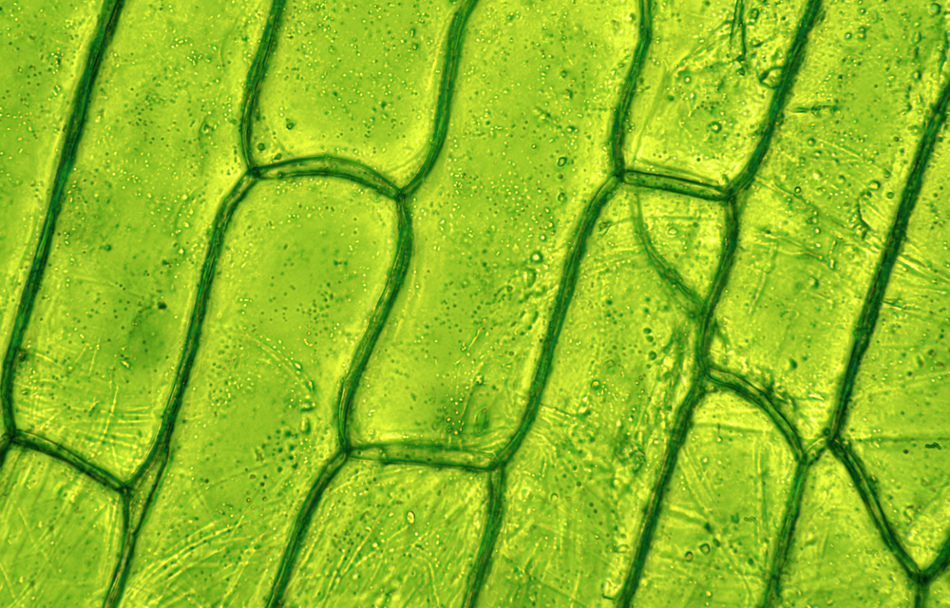
Image Credit: Barbol/Shutterstock.com
Microscopy such as transmission electron microscopy (TEM) and field emission scanning electron microscopy (FESEM) have been instrumental for the study of the structure of cell walls. However, these techniques require fixed material, which is not always representative of native hydrated cell walls. The evolution in microscopy, for example, fluorescence microscopy, makes it possible for the detailed study of the cell wall and various plant protein interactions.
Why is Research on Plant Cell Walls Important?
Scientists at STFC’s Central Laser Facility (CLF) revealed that plant cell walls play a vital role in regulating plant protein trafficking than previously understood. The research on cell walls led by Dr. Joseph McKenna from Oxford Brookes University revealed that cell walls regulate an extraordinary number of plant functions through proteins trafficking within the cell. Their research shows that manipulating the cell walls could result in higher productivity in crops with greater resistance to disease.
The team of cell biologists at Oxford Brookes University studied the interaction between proteins and cell walls in plants. In their study, they found how proteins move hormones between cells, which is indirectly responsible for plant growth.
Dr. Dan Rolfe, Lead Data Scientist at Octopus, investigated a single molecule tracking system where a thin layer of the surface of the cell is illuminated with the laser to visualize individual or small numbers of molecules. This allows researchers to trace the molecular paths in cells. Therefore, laser microscopy is useful as it can reveal finer details in the behaviors of particular molecules.
Microscopic Techniques Used to Analyze Cell Wall and Plant Protein Interaction
Various high-resolution techniques have recently emerged that show great promise for cell wall research, often containing several strengths and weaknesses. Super-resolution microscopy includes multiple techniques that help overcome diffraction limitations that affect the resolution in classical far-field optical microscopy systems.
Resolution in conventional far-field systems is directly proportional to the wavelength of excitation light and inversely proportional to the numerical aperture of the light-collecting lens.
The first major category of super-resolution microscopy includes techniques that illuminate the sample by fixed patterned light (linear and nonlinear), such as structured illumination microscopy (SIM) and stimulated emission depletion microscopy (STED).
The second main category of super-resolution techniques includes single-molecule localization strategies that analyze the positioning of individual fluorophores with sub-diffraction accuracy. Such methods mainly involve the following:
- Photoactivation localization microscopy (PALM)
- Fluorescence photoactivated localization microscopy (FPALM)
- Stochastic optical reconstruction microscopy (STORM)
- Related variants such as super-resolution optical fluctuation microscopy (SOFI)
Laser scanning confocal microscopy (LSCM) and spinning disk confocal microscopy (SDCM) are used to study the dynamics of proteins involved in cell wall synthesis. LSCM uses a single pinhole for optical sectioning. However, SDCM uses an array of excitation and emission pinhole apertures on a rapidly spinning disk, such that the pinhole array sweeps the entire field of view over 1,000 times per second.
Click here to find out more about the different types of microscopes.
The high scan speed improves image acquisition rate and lowers the peak excitation light density down to a few μW/μm2. This increases fluorescence efficiency and decreases photobleaching and photodamage effects compared to point scanning.
Fluorescence recovery after photobleaching (FRAP) and fluorescence loss in photobleaching (FLIP) are useful complementary tools for imaging cell wall proteins. When using a confocal microscope, the yellow fluorescent protein appears as transverse bands in developing xylem vessels.
The intracellular trafficking of proteins can be appropriately examined using photoactivatable or photoconvertible fluorescent proteins. Photoactivatable fluorescent proteins are used in plant science research to analyze the relationship between the Endoplasmic Reticulum and Golgi stacks in tobacco leaf epidermal cells or the distribution of KAT1 K+ channels in tobacco leaves. The property of photoconvertible fluorescent proteins to show pronounced light-induced spectral changes allows pulse-chase analysis of protein trafficking.
Total internal reflection fluorescence microscopy (TIRFM) is used for the detection of molecules at the surface of plant cells. This technique involves total internal reflection (TIR) that occurs when a ray of light strikes a boundary between two materials with different refractive indices (n) and the incident angle is greater than the critical angle of incidence. During these conditions, all of the light is reflected into the medium with a higher n value. When TIR occurs, an evanescent wave (EW) forms at the boundary, which penetrates the surface of the medium to a depth approximately equal to one-third of the wavelength of the incident light.
A TIRF-derived method, known as variable-angle epifluorescence microscopy (VAEM) has recently been developed to visualize vesicle trafficking and fusion events at the plasma membrane in plant cells.
Key Organizations Manufacturing Advanced Microscopes
Several microscopic systems are established by different research groups as well as commercial companies. Some of the prominent companies that make different kinds of advanced microscopes are listed below:
- Nikon (N-STORM; N-SIM; combination of N-SIM and N-STORM)
- Leica Microsystems (Leica SR GSD 3D microscope – STORM, Leica TCS SP8 STED 3X - STED)
- Carl Zeiss (Elyra P.1 - PALM; Elyra S.1 - SIM; Elyra PS.1 - combination of SIM and PALM)
- GE Healthcare Life Sciences (DeltaVision Localization Microscopy System - STORM; DeltaVision OMX System - SIM; combination of SIM and STORM)
References and Further Reading
Gonneau, M., Höfte, H. and Vernhettes S. (2012). Fluorescent tags to explore cell wall structure and dynamics. Frontiers in Plant Science. 3,145.
McKenna J. F., Rolfe, D. J. et al. (2019). The cell wall regulates dynamics and size of plasma-membrane nanodomains in Arabidopsis. Proceedings of the National Academy of Sciences. 116 (26) 12857-12862.
Komis, G., Novák, D. et al. (2018). Advances in Imaging Plant Cell Dynamics. Plant Physiology. 176, 80–93.
The Science and Technology Facilities Council on behalf of STFC (2019) Lasers Reveal the Secrets of Plant Cell Walls. [Online] Labmate. Available at: https://www.labmate-online.com/article/microscopy-and-microtechniques/4/stfc/lasers-reveal-the-secrets-of-plant-cell-walls/2647 (Accessed on 5 May 2020).
Disclaimer: The views expressed here are those of the author expressed in their private capacity and do not necessarily represent the views of AZoM.com Limited T/A AZoNetwork the owner and operator of this website. This disclaimer forms part of the Terms and conditions of use of this website.