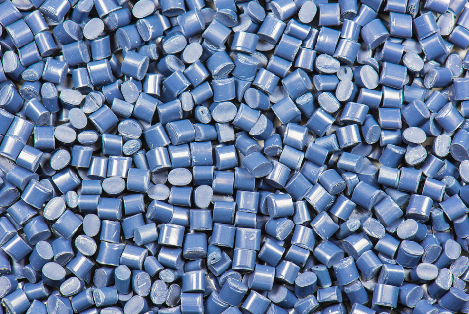
Image Credit: XXLPhoto/Shutterstock.com
The use of microscopic techniques for the analysis of synthetic polymers is incredibly useful in providing researchers with detailed information on the rheological, mechanical, optical, electrical and/or chemical properties of these materials.
Microscopy of Polymers
The structural analysis of polymers can be achieved using a variety of analytical instruments; however, various microscopy techniques have proven to be indispensable for this purpose. Some of the most common microscopy tools used within the polymer industry include light, electron, confocal and polarization microscopy. These instruments are often used to assess the structure, fracture planes, roughness, strain, crystal morphology and spherulites of polymer samples.
The failure analysis of failure polymer parts or products is often achieved through the use of light microscopy. By analyzing the surface structure of fraction planes, light microscopy images can elucidate any potential causes associated with the polymer’s failure and/or defects, as well as the origin of any detected cracks.
Similarly, applications that require surface topography analyses of polymers often turn to confocal microscopy. Whereas upright and polarization microscopy is typically used to assess the mechanical, biodegradable and biocompatible products of polymers, electron microscopy (EM) is instead used during several different polymer preparation processes.
High-Resolution Optical Microscopes
Traditional optical microscopy techniques were often limited in their ability to accurately characterize the structural and dynamic properties of synthetic polymers because of their low spatial resolution.
The recent development of high-resolution optical microscopes, such as superlocalization microscopy (SLM), spectrally resolved super-resolution microscopy and stimulated emission depletion (STED) microscopy have overcome such issues, opening the door for these techniques to be applied to synthetic polymer characterization.
Click here for more information on optical microscopes.
The information obtained by these methods has been found to be complementary to that obtained by a number of other characterization methods such as EM and scanning probe microscopy, as well as both X-ray and neutron scattering. In fact, these novel optical microscopy techniques have been found to offer certain advantages over to these other characterization methods, without compromising on acquired information.
For example, while EM may offer a greater spatial resolution for morphological analyses, this method can only measure dried samples under vacuum conditions, limiting the type of samples that can be analyzed. By eliminating this concern, high-resolution optical microscopy techniques are able to investigate the spatiotemporal heterogeneity of polymer materials’ properties while also achieving a similar resolution to that of EM.
SLM
Several novel SLM methods have been successfully applied to work investigating the unique properties of synthetic polymers. For example, a recent study utilized SLM for the characterization of hydrogel-based nanostructures that had been previously immobilized on substrate surfaces.
Another study found that SLM provided a unique insight into the chemical characterization of polymer surfaces, as it was able to visualize the distribution of carboxylic acid groups present on two different thermoplastic surfaces. In fact, the results of this study determined SLM to be a superior imaging method compared to conventional fluorescence microscopy for this purpose.
STED
A recent study described how a modified STED microscopy method successfully performed subdiffraction-limited imaging of conducting polymer nanoparticles (CPNs). In this work, the traditional STED method altered the excitation beam of this instrument to allow for a synchronous detection of two-photon-excited fluorescence background, improving the resolution of capturing CPNs.
Another study utilized a STED-type microscope found that this instrument could map the local intensity of electroluminescence in an effort to detect defects present within the samples.
Conclusion
Overall, high-resolution microscopy techniques have been found to be highly versatile tools that can be manipulated to assess specific polymer properties. As more work emerges in this area, researchers are confident that optical methods will be further adjusted to allow for nanoscale analysis of polymers found in surface coating, chemical separation, chemical sensing, bioimaging and organic electronic devices.
References and Further Reading
Disclaimer: The views expressed here are those of the author expressed in their private capacity and do not necessarily represent the views of AZoM.com Limited T/A AZoNetwork the owner and operator of this website. This disclaimer forms part of the Terms and conditions of use of this website.