 By Owais AliReviewed by Lexie CornerSep 27 2024
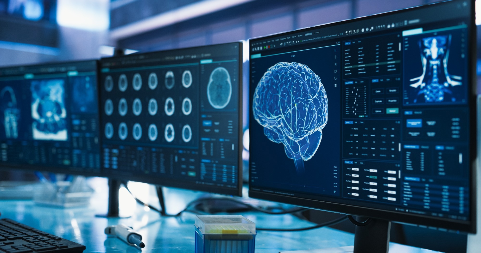
Brain imaging techniques are used to visualize and map changes in brain structure and function related to clinical disorders, offering insights into the underlying molecular mechanisms. This article reviews recent advancements in brain imaging techniques and their significance in enhancing diagnostic and therapeutic approaches.

Image Credit: Gorodenkoff/Shutterstock.com
The human brain regulates all involuntary and voluntary movements, as well as complex cognitive functions critical for high-level living. With rising life expectancy, the prevalence of brain disorders, including age-related and social diseases, has increased, often worsened by environmental changes, social stress, and addictions.
Despite advancements in neurological treatments, the prognosis for these disorders remains poor due to the brain's susceptibility to microdamage, highlighting the need for highly resolved, rapid, and non-invasive imaging techniques for accurate diagnosis.
Current Brain Imaging Technologies
Electroencephalography (EEG)
In 1924, German psychiatrist Hans Berger developed the first-ever brain imaging technique: the EEG.1 It measures brain waves using hundreds of electrodes attached to the scalp with special glue or a cap. When neurons fire, they generate electrical fields, which are recorded as wave patterns that can appear as random squiggly lines.
EEG analysis focuses on five primary wave patterns: gamma waves (active learning), beta waves (awake but relaxed), alpha waves (relaxed or daydreaming), theta waves (deep thought), and delta waves (deep sleep).
Neurologists assess these patterns for abnormalities, enabling the diagnosis of neurological and mental disorders, including epilepsy, brain tumors, brain injuries, strokes, and sleep disorders.2,3
Magnetic Resonance Imaging (MRI) and Functional MRI (fMRI)
MRI is a non-invasive technique that generates detailed three-dimensional images of the brain and body using magnetic fields and radio waves to manipulate hydrogen protons in tissues. This process creates cross-sectional images through radiofrequency pulses, making it particularly effective for imaging hydrogen-rich soft tissues and tracking dynamic changes in the brain over time.
fMRI builds on this by indirectly measuring neural activity through changes in blood flow, using the blood-oxygen-level dependent (BOLD) contrast method. It provides high spatiotemporal resolution, enabling precise functional brain mapping during various cognitive tasks and in the study of neural disorders. fMRI is widely employed in brain mapping and is instrumental in the diagnosis and surgical planning for conditions such as epilepsy, brain gliomas, schizophrenia, and Alzheimer's disease.2-4
Positron Emission Tomography (PET)
PET is another imaging modality that provides insights into tissue functionality. It utilizes radioactive tracers that bind to glucose, highlighting areas of the brain with high metabolic activity.
PET scans create images where active regions are indicated by warmer colors, reflecting varying neuronal activity levels.
Compared to MRI, PET is superior for measuring metabolic activity, particularly in identifying changes associated with neurodegenerative diseases, such as Alzheimer's.2,4,5
Computed Tomography (CT)
CT scanning employs specialized X-ray technology to generate detailed cross-sectional brain images. The CT scanner rotates around the body, capturing data from multiple angles to create two-dimensional images that can be compiled into three-dimensional representations for comprehensive analysis.
It offers superior clarity compared to standard X-rays and can utilize contrast agents to enhance visualization of blood vessels and tumors, making it valuable for diagnosing injuries, diseases, and conditions such as brain hemorrhages and tumors.5,6
Limitations of Traditional Brain Imaging Technologies
While these traditional brain imaging technologies offer valuable insights, they have significant limitations that can impact their effectiveness and safety.
- EEG primarily detects activity in superficial cortical layers, lacking sensitivity to deeper brain structures. Its low spatial resolution complicates the reconstruction of intracranial current sources.
- fMRI has poor temporal resolution due to the slow BOLD response, which indirectly measures neuronal activity and is influenced by unrelated physiological processes, complicating interpretations of brain function.
- The noise from the detection of photons affects PET, impacting spatial resolution and overall imaging clarity.
- CT scans expose patients to ionizing radiation, which can pose significant risks, especially for pediatric patients. Additionally, the use of contrast agents may cause allergic reactions and contrast-induced nephropathy in susceptible individuals.
Recent Technological Advances and Their Impact on Neuroscience research
AI-Enhanced Susceptibility Tensor Imaging (STI)
STI is an advanced MRI technique that employs a second-order tensor model to characterize anisotropic tissue magnetic susceptibility. It offers insights into white matter fiber pathways and myelin alterations in the brain with millimeter-scale resolution, enhancing understanding of brain structure and function in healthy and diseased states.
Recently, John Hopkins University researchers developed a DeepSTI algorithm, which significantly optimizes this process by generating high-resolution three-dimensional maps of brain tissue with fewer MRI scans than traditional STI methods. This algorithm employs machine learning techniques and regularization approaches that concentrate on the most plausible reconstructions of brain images, enhancing the clarity and accuracy of the resulting data.
The DeepSTI algorithm allows for detailed brain scans using only one head orientation, reducing the procedure's duration and increasing patient comfort. This method enables visualization of critical tissue changes, like myelin alterations in MS patients, facilitating better disease monitoring.
The improved efficiency and quality of STI imaging are expected to enhance its clinical applicability, aiding in the diagnosis and treatment planning for various neurological disorders.7
Dual Imaging Techniques for Mapping Neural Connectivity and Microstructure
Diffusion MRI (dMRI) is a non-invasive technique that utilizes the diffusion of water molecules to elucidate structural connectivity, proving especially valuable for tumor characterization and evaluating cerebral ischemia. This method enhances the understanding of tissue pathology and informs treatment strategies.
Polarization-sensitive optical coherence tomography (PS-OCT) complements this by employing back-scattered light and polarization variations to produce depth-resolved cross-sectional images.
A recent study compared nerve fiber orientations in the human brainstem using PS-OCT and dMRI-based tractography. The research highlighted that while dMRI provides insights into structural connectivity, it cannot detect specific cellular changes. In contrast, PS-OCT offers micrometer-scale resolution for identifying fiber tracts and distinguishing between white and gray matter, though it is limited by depth in scattering media.
Therefore, PS-OCT can validate dMRI results, enhancing understanding of nerve fiber microstructure critical for normal physiology and neurodegenerative disease insights.8
Investigating Blood Flow Dynamics and Neuronal Activity with fUSI
Functional ultrasound imaging (fUSI) uses highly sensitive ultrasound to measure blood volume changes at the capillary level, indirectly indicating neuronal activity. This fast, safe, and portable method enables deep tissue imaging in awake or freely moving subjects with high spatiotemporal resolution (100 ms, 100 μm) and a wide field of view covering the entire rodent brain.
These attributes make fUS ideal for investigating large-scale brain integration and specific neuronal circuits, while its high resolution and portability enhance its potential for clinical applications.9
In a study published in the Annual Review of Neuroscience, researchers implanted a transparent window in a patient's skull and used fUSI to obtain high-resolution brain imaging data. This clear implant allows for detailed functional data acquisition related to blood flow and electrical activity, essential for understanding normal brain function and various neurological conditions.
The results indicated that fUSI could effectively gather functional imaging data through the implant, offering insights into nerve fiber activity and potentially aiding in diagnosing and treating conditions such as epilepsy and dementia.
This approach presents a promising alternative to traditional methods like fMRI and intracranial EEG, which often require invasive procedures.10
Conclusion
Recent advancements in brain imaging technologies have significantly improved the understanding of neuronal activity and structural connectivity, enhancing diagnostic and therapeutic strategies for neurological conditions.
Current research is dedicated to refining these techniques and broadening their applications, with a focus on non-invasive methods that have the potential to transform clinical practices and provide deeper insights into brain function.
Discover More: Neuroplasticity: Rewiring the Brain
References and Further Reading
- Britton, JW., et al. (2016). Electroencephalography (EEG): An introductory text and atlas of normal and abnormal findings in adults, children, and infants. American Epilepsy Society. https://www.ncbi.nlm.nih.gov/books/NBK390348/
- Bosquez, T. (2022). Neuroimaging: Three important brain imaging techniques. [Online] The Indiana University Bloomington. Available at: https://blogs.iu.edu/sciu/2022/02/05/three-brain-imaging-techniques/
- Xue, G., Chen, C., Lu, ZL., Dong, Q. (2010). Brain Imaging Techniques and Their Applications in Decision-Making Research. Xin li xue bao. Acta psychologica Sinica. https://doi.org/10.3724/SP.J.1041.2010.00120
- Kim, B., et al. (2021). A brief review of non-invasive brain imaging technologies and the near-infrared optical bioimaging. Appl. Microsc. https://doi.org/10.1186/s42649-021-00058-7
- Vaquero, JJ., Kinahan, P. (2015). Positron Emission Tomography: Current Challenges and Opportunities for Technological Advances in Clinical and Preclinical Imaging Systems. Annual review of biomedical engineering. https://doi.org/10.1146/annurev-bioeng-071114-040723
- Patel PR, De Jesus O. (2023). CT Scan. [Online] National Library of Medicine. Available from: https://www.ncbi.nlm.nih.gov/books/NBK567796/
- Fang, Z., Lai, KW., van Zijl, P., Li, X., Sulam, J. (2023). Deepsti: towards tensor reconstruction using fewer orientations in susceptibility tensor imaging. Medical image analysis. https://doi.org/10.1016/j.media.2023.102829
- Optica. (2024). Integrating MRI and OCT for new insights into brain microstructure. [Online] Frontiersinoptics. https://www.frontiersinoptics.com/home/media-center/conference-news/integrating-mri-and-oct-for-new-insights-into-brai/
- Montaldo, G., Urban, A., Macé, E. (2022). Functional ultrasound neuroimaging. Annual Review of Neuroscience. https://doi.org/10.1146/annurev-neuro-111020-100706
- Rabut, C., Norman, SL., Griggs, WS., Russin, JJ., Jann, K., Christopoulos, V., Liu, C., Andersen, RA., Shapiro, MG. (2024). Functional ultrasound imaging of human brain activity through an acoustically transparent cranial window. Science translational medicine. https://doi.org/10.1126/scitranslmed.adj3143
Disclaimer: The views expressed here are those of the author expressed in their private capacity and do not necessarily represent the views of AZoM.com Limited T/A AZoNetwork the owner and operator of this website. This disclaimer forms part of the Terms and conditions of use of this website.