Up until now, measuring with high sensitivity and high spatial resolution with X-Ray light in the delicate energy range of 1.5–5.0 keV has been incredibly laborious. However, this X-Ray light is excellent for examining biological systems and energy-producing components like batteries or catalysts.

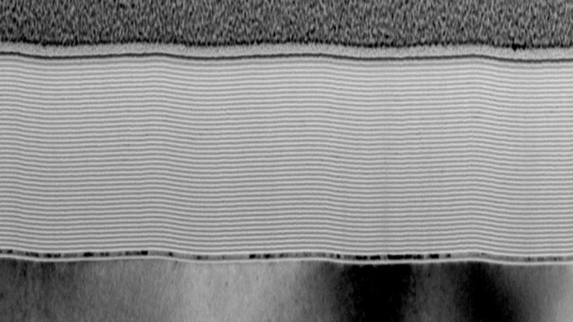
Schematic drawing of the novel monochromator concept at the U41-PGM1 beamline at BESSY-II based on a multilayer coated blazed plane grating and mirror to improve the photon flux in the tender X-ray photon energy range (1.5 – 5.0 keV). The inset shows a TEM image of the cross-section of the Cr/C multilayer blazed grating structures. For better visualization of the grating period, the image was horizontally compressed 10 fold. Image Credit: HZB / Small Methods 2022
The newly created monochromator optics boost the photon flux in the tender energy range by a factor of 100, enabling extremely accurate measurements of nanostructured systems, according to a team from HZB, which has now found a solution to this issue. On catalytically active nanoparticles and microchips, the technique was successfully tested for the first time.
A wide range of materials, such as catalytically active materials and new battery electrodes, are needed for energy conversion processes to produce a supply of energy that is climate neutral. The presence of nanostructures in many of these materials improves their functionality.
When analyzing these samples, high-resolution nanoscale X-Ray imaging and spectroscopic measurements to identify the chemical properties work best together.
But up until now, there has been a significant issue because important components of these materials, like molybdenum, silicon, or sulfur, primarily react to X-Rays in the so-called tender photon energy range.
This is due to the fact that conventional X-Ray optics from a plane grating or crystal monochromators only deliver very low efficiencies in this “tender” energy range between soft and hard X-Rays. This issue has now been resolved by the HZB team.
We have developed novel monochromator optics. These optics are based on an adapted, multilayer-coated sawtooth grating with a plane mirror.
Frank Siewert, Senior Scientist, Department Optics and Beamlines, Helmholtz-Zentrum Berlin
The new monochromator concept multiplies the photon flux in the sensitive X-Ray range by 100, enabling for the first time extremely sensitive spectromicroscopic measurements with high resolutions.
Within a short time, we were able to collect data from NEXAFS spectromicroscopy on the nanoscale. We have demonstrated this on catalytically active nanoparticles and modern microchip structures. The new development now enables experiments that would otherwise have required months of data collection.
Stephan Werner, Study First Author and Physicist, Department X-Ray Microscopy, Helmholtz-Zentrum Berlin
Gerd Schneider, who heads the X-Ray Microscopy Department at HZB, stated, “This monochromator will become the method of choice for imaging in this X-ray energy range, not only at synchrotrons worldwide, but also at free-electron lasers and laboratory sources.”
He anticipates profound impacts on a wide range of material research areas, including studies in the sensitive X-Ray range that could significantly advance the development of energy materials and thereby help to develop climate-neutral solutions for the production of electricity and other forms of energy.
Journal Reference
Werner, S., et al. (2022) Spectromicroscopy of Nanoscale Materials in the Tender X-Ray Regime Enabled by a High Efficient Multilayer-Based Grating Monochromator. Small Methods. doi:10.1002/smtd.202201382.