Particulate dispersions flow inside the anterior chamber of the human eye due to a variety of reasons. These scattered intraocular particles can impair vision. Intraocular particles also increase pressure levels inside the eye resulting in secondary issues such as particle-related glaucoma, a leading cause of blindness.

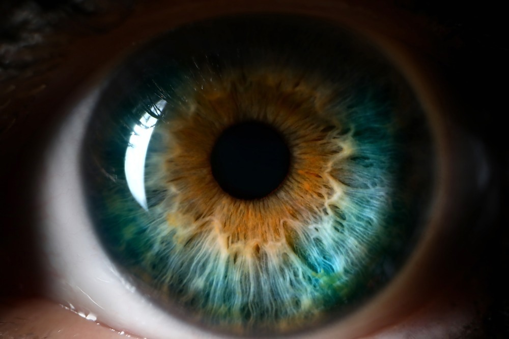
Study: Acoustic Manipulation of Intraocular Particles. Image Credit: H_Ko/Shutterstock.com
Theoretically, these issues can be avoided by managing these scattered particles in a way that lessens their detrimental effects. However, it is challenging to manipulate intraocular particles inside the eye.
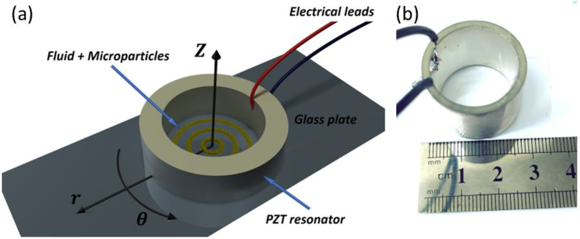
A recent study published in Micromachines describes a brand-new technique that uses acoustic waves to alter intraocular particles without entering the anterior chamber. The researchers demonstrate ex vivo modulation of pigmentary particles in swine eyes using a cylindrical sonic resonator.
The researchers examine how wave intensity changes over time and rule out any temperature changes that would cause tissue injury. This technique can regulate and arrange intraocular particles in particular regions without harming ocular tissue and allow regular aqueous humor outflow, which is essential for preserving the right intraocular pressure levels.

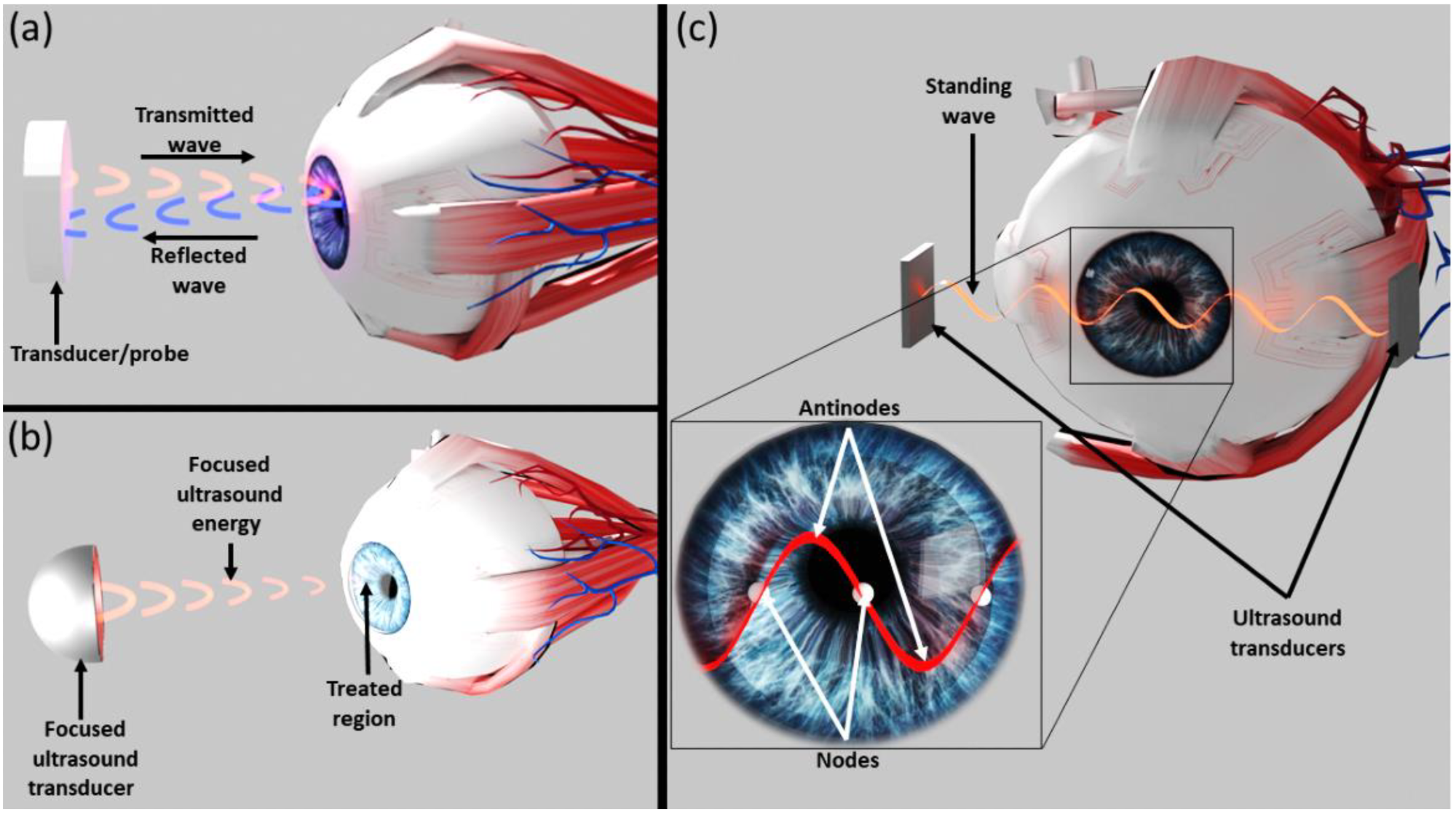
Figure 1. Illustration of various methods that utilize acoustic waves in ophthalmology. (a) Ultrasonic waves used to image the location and characteristics of various eye components. (b) High-intensity focused ultrasound used to destroy ocular tissue ablation. (c) The novel method for interocular particle acoustic manipulation suggested in this manuscript. Acoustic transducers are used to create standing acoustic waves that direct intraocular particles towards their nodal areas. © Leshno, A., Kenigsberg, A., Peleg-Levy, H., Piperno, S., Skaat, A., & Shpaisman, H. (2022)
Clinical Significance of Aqueous Humor (AH)
The ciliary body secretes a complex mixture of biological molecules into the posterior chamber of the eye to form aqueous humor (AH), a clear, low-viscosity fluid. The humor then moves to the anterior chamber before draining into the systemic circulatory system. The trabecular meshwork (TM), corneal endothelium, and lens are three nearby non-vascularized tissues that require AH circulation to survive and be protected.
Secretion and outflow control of AH are crucial physiological processes for the standard operation of the human eye. Average intraocular pressure required to preserve the normal shape and optical qualities of the healthy eye is maintained by the flow of AH against resistance. Reduced AH outflow and decreased AH transparency result in reduced visual acuity and increased intraocular pressure, a major cause of glaucoma and other ocular diseases. Synthesis, drainage, circulation, and consistency of AH are of significant therapeutic importance.
Negative Impacts of Dispersion of Particulate Matter on AH
Particulate matter of various sizes, shapes, and features can develop or disperse under a variety of circumstances and circulate into the AH. These particles can restrict the TM and the AH outflow, leading to secondary problems including open-angle glaucoma and obstructing the visual axis in the acute phase. Intraocular particles can settle in the corneal endothelium and cause Krukenberg's spindle or long-lasting hyphema corneal staining.
Although there are surgical and medicinal treatments available for these issues, good preventative measures are lacking. The current clinical practice largely adopts a "wait and see" strategy, treating only secondary issues and providing no options for disposing of particulate matter.
Utilization of Sound Waves in Medical Examination
Sound waves with auscultation are employed in physical examination. The most widespread use of acoustics in medicine is ultrasonography. Ultrasonography uses sound waves with frequencies higher than the upper limit of human hearing and is utilized for imaging.
An acoustic transducer used in ophthalmology generates waves reflected by different eye parts to produce images. By monitoring the relative amplitudes and times of the echoes, a probe (or, optionally, the emitting transducer) can determine the position and properties of the reflecting components.

Figure 2. (a) Illustration (not to scale) of the experimental in vitro apparatus. The dispersed micro particles are inserted into the piezoelectric resonator. They are driven towards nodal areas and form rings upon activation of the acoustic waves. (b) Photograph of the in vitro apparatus. © Leshno, A., Kenigsberg, A., Peleg-Levy, H., Piperno, S., Skaat, A., & Shpaisman, H. (2022)
Development of Acoustic Manipulation to Control Intraocular Particles
Leshno et al. proposed using acoustic manipulation to regulate intraocular particles with potential clinical implications to manage and prevent problems caused by particulate matter. The approach uses acoustic manipulation to control particle flow within the AH by limiting tissue interaction with particles and eliminating the need for surgical intervention.
A series of techniques known as acoustic manipulation use acoustic radiation forces to move particles. When a sound field is applied to a fluid dispersion containing solid particles, those particles either scatter or absorb the sound field. Intraocular particles can be moved to specified points in space where they can cluster by certain wave properties.
Acoustic manipulation techniques have a significant advantage over other methods like optical, optoelectronic, electric, and magnetic tweezers because their only prerequisite is a difference in density and compressibility between the particles present in almost all dispersed systems. Additionally, the power density needed to control intraocular particles using acoustic manipulation forces is several orders of magnitude lower than their optical counterparts and has a far wider field of action.

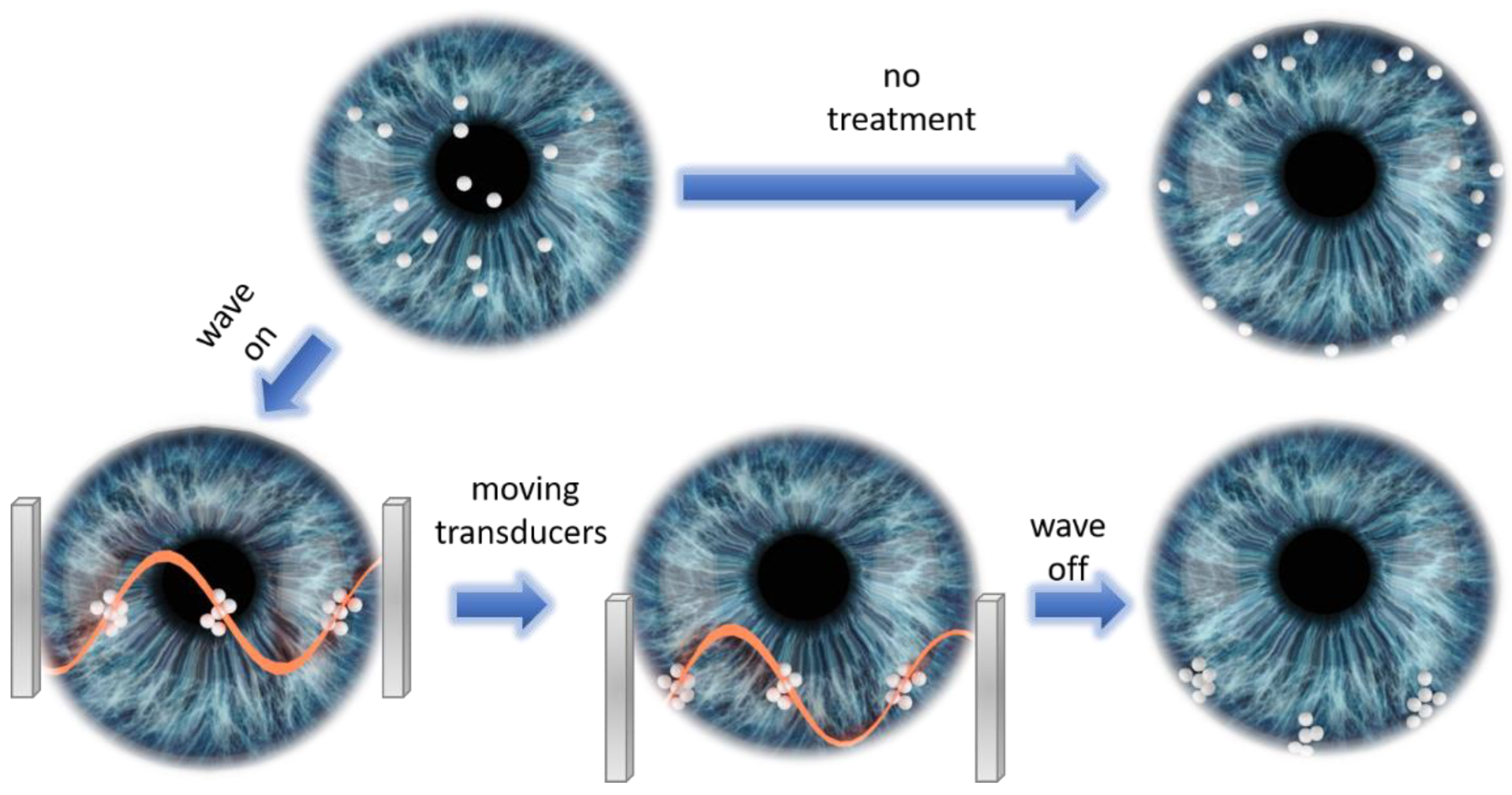
Figure 3. Illustration of suggested protocol utilizing acoustic manipulation for preventing secondary glaucoma. Without treatment, particles will flow towards the angle and distribute uniformly on the TM. Acoustically trapping particles at nodal areas followed by moving the acoustic transducers leads to placement of trapped particles at specific locations of the angle, thus leaving unaffected areas of TM that can support AH outflow. © Leshno, A., Kenigsberg, A., Peleg-Levy, H., Piperno, S., Skaat, A., & Shpaisman, H. (2022)
Research Findings
This investigation showed that the mobility of particles within the anterior chamber could be controlled using acoustic manipulation forces without causing any harm to the cornea. The main advantage of the suggested technique is the viability and safety of using sonic manipulation techniques to noninvasively control particle mobility within the anterior chamber without harming ocular tissue.
It is the first stage in creating ocular acoustic systems, which will enable more advanced diagnoses and therapies for conditions where there are currently no available remedies. The ability to move and manipulate the particles noninvasively is a key benefit of the method, as it lessens concerns about consequences from intraocular intervention. More research is necessary to understand this unique technology's capabilities and potential applications in ophthalmology.
Reference
Leshno, A., Kenigsberg, A., Peleg-Levy, H., Piperno, S., Skaat, A., & Shpaisman, H. (2022). Acoustic Manipulation of Intraocular Particles. Micromachines, 13(8), 1362. https://www.mdpi.com/2072-666X/13/8/1362/htm.
Disclaimer: The views expressed here are those of the author expressed in their private capacity and do not necessarily represent the views of AZoM.com Limited T/A AZoNetwork the owner and operator of this website. This disclaimer forms part of the Terms and conditions of use of this website.