A ground-breaking technique formulated by the Prevedel Group at European Molecular Biology Laboratory (EMBL) enables neuroscientists to track live neurons deep inside the brain — or any other cell concealed within an opaque tissue.
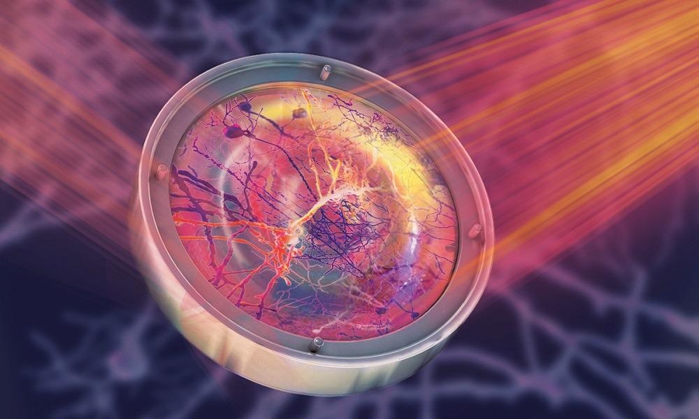
 A deformable mirror used in microscopy to focus light within live tissues. They would normally distort its propagation. Thanks to this mirror, we can see clear and crisp images of neuronal cells deep inside the brain. Image Credit: Isabel Romero Calvo/EMBL.
The novel approach combines two advanced microscopy methods, adaptive optics, and three-photon microscopy. The September 30th issue of Nature Methods reports about this advancement.
Before the development of the new method, neuroscientists found it challenging to monitor astrocytes producing calcium waves in the deep layers of the cortex or to envisage any other neural cells in the hippocampus, a region deep in the brain that is in control of navigation and spatial memory. The phenomenon occurs recurrently in the brains of all live mammals.
By formulating the new method, Lina Streich from the Prevedel Group and her collaborators could capture minute details of these versatile cells at unparalleled high resolution. The international team was made up of scientists from Austria, Argentina, India, China, Germany, France, the United States and Jordan.
In the neurosciences, brain tissues are analyzed typically in small model organisms or in ex vivo samples that have to be sliced up to be analyzed — both of which signify non-physiological situations. Usual brain cell activity occurs only in live animals, but the “mouse brain is a highly scattering tissue,” said Robert Prevedel.
In these brains, light cannot be focused very easily, because it interacts with the cellular components. This limits how deep you can generate a crisp image, and it makes it very difficult to focus on small structures deep inside the brain with traditional techniques.
Robert Prevedel, Physicist, Prevedel Group, EMBL
According to Streich, a former PhD student in the lab who tackled this issue for more than four years, researchers can now observe further into tissues.
With traditional fluorescence brain microscopy techniques, two photons are absorbed by the fluorescence molecule each time, and you can make sure that the excitement caused by the radiation is confined to a small volume. But the further the photons travel, the more likely they are lost due to scattering.
Robert Prevedel, Physicist, Prevedel Group, EMBL
One approach to surpass this is to expand the wavelength of the exciting photons toward the infrared, which guarantees that the fluorophore absorbs sufficient radiation energy. Furthermore, using three photons rather than two enables clearer images deep within the brain to be obtained. But another hurdle remains: ensuring that the photons are concentrated so that the entire image is not hazy.
This is where the second method applied by Streich and her team is crucial. Adaptive optics is used frequently in astronomy — and without a doubt, it was pivotal for Reinhard Genzel, Roger Penrose and Andrea Ghez to win the 2020 Nobel Prize in Physics for their discovery of black holes.
To correct the distortion in the light wavefront triggered by atmospheric turbulence, astrophysicists use deformable, computer-regulated mirrors to rectify in real-time. In Prevedel’s lab, the distortion is caused by the scattering of inhomogeneous tissue, but the technology and the principle are quite similar.
“We also use an actively controlled deformable mirror, which is capable of optimising the wave fronts to allow the light to converge and focus even deep inside the brain,” described Prevedel.
“We developed a custom approach to make it fast enough to use on live cells in the brain,” added Streich.
To minimize the invasiveness of the method, the team also reduced the number of measurements required to attain high-quality images.
This is the first time these techniques have been combined and thanks to them, we were able to show the deepest in vivo images of live neurons at high resolution.
Lina Streich, Prevedel Group, EMBL
The researchers, who worked in partnership with colleagues from EMBL Rome and the University of Heidelberg, even observed the dendrites and axons that link the neurons in the hippocampus, while leaving the brain totally intact.
“This is a leap towards developing more advanced non-invasive techniques to study live tissues,” Streich stated.
Although the method was designed for use on a mouse brain, it is readily applicable to any opaque tissue.
Besides the obvious advantage of being able to study biological tissues without the need to sacrifice the animals or to remove overlaying tissue, this new technique opens the way to study an animal longitudinally, that is, from the onset of a disease to the end. This will give scientists a powerful instrument to better understand how diseases develop in tissues and organs.
Lina Streich, Prevedel Group, EMBL
Journal Reference:
Streich, L., et al. (2021) High-resolution structural and functional deep brain imaging using adaptive optics three-photon microscopy. Nature Methods. doi.org/10.1038/s41592-021-01257-6.