Apr 10 2017
ChipScope is a European project led by the University of Barcelona with the participation of SMEs, research institutes, and universities from five European countries. The project focuses on developing a new type of chip-sized optical microscope with high resolution capabilities. The aim is to develop the required science and technology to observe minute structures such as DNA molecules, viruses, or the inside of the cells, in real-time by avoiding the limitations of the existing high resolution techniques. The project has been scheduled for a 4-year length period and is funded with 3.75 million euros by Future and Emerging Technologies (FET Open), which is a program focused on research to develop breakthrough technologies.
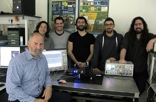
 The project ChipScope will be conducted between January 2017 and December 2020, led by the University of Barcelona and coordinated by Angel Dieguez. Credit: University of Barcelona
ChipScope will be conducted from January 2017 and December 2020, and it is led by the University of Barcelona, with the participation of the Braunschweig University of Technology (Germany), the University of Rome Tor Vergata, the company Expert Ymaging (Barcelona), the Austrian University of Technology, the Medical University of Vienna, and the Swiss Foundation for Research in Microtechnology.
Overcoming the limits of diffraction
200 nm is the minimum distance needed to distinguish two independent elements with a microscope. In other words, it is about five hundred times smaller than human hair. DNA molecules, proteins, or internal cell structures are smaller than this size, and thus, cannot be directly observed by using common optical microscopes.
At the moment, these kind of observations under the called diffraction limit are only possible with complex and expensive electronic microscope which, in addition, end up destroying the sample
Ángel Diéguez, member of the Research Group of Instrumentation Systems and Communication (SIC) of the UB and coordinator of the project.
ChipScope aims to develop a new type of miniature microscope that will enable samples to be observed below the diffraction limit, without altering the sample. For this purpose, researchers will adopt a technique different from the one adopted for the common microscopes: “The idea is that the resolution has to depend more on the light source than the optical detection system. That is, instead of an only source of light, like it happens with current high resolution microscopes, we will use hundreds of miniature light sources”, says Ángel Diéguez, whose group is well-versed with the development of miniature microcircuits.
World’s smallest LEDs
The new technique will involve the fabrication of the world’s smallest LEDs (about 50 nm in size), which will act as light sources for the new microscope. These nano-LEDs will be uniformly spaced apart at nanometric distances in a matrix that will form the basis for the new tool. When the nano-LEDs are individually switched on at a high speed, it will enable the user to determine which information is originating from which position of the observed object. These signals will be detected by a highly sensitive photodetector, so that a real-time image of the object can be transferred.
The theoretical base of the project was already thought of in the sixties, but in order to work on those ideas, they needed microchips, LEDs and the ability to build those nanometric-sized objects and place them in order.
Daniel Prades, member of the Research Group Micro-and-nanotechnologies for Electronics and Photonics (MIND), which is also part of this project.
Technology to set new science up
Developing a microscope with the above-said features will pave the way for new opportunities in scientific research, both for the advancement in technological miniaturization as well as for the new physical effects to be studied in order to carry out the project. Additionally, this is a technology that will allow — once it is completed — “to imagine new experiments”.
Having a microscope of this size will provide the opportunity of measuring things in conditions that were impossible so far. This technology will set new science up, since it will enable creating new experiments, such as observing places you could never get into with an optical microscope.
Prades.
The initial tests with the new microscope will be conducted on cell samples of Idiopathic Lung Fibrosis (ILF), a chronic age-related lung disease. ILF affects humans and causes 0,5 million deaths annually.