Researchers at Massachusetts General Hospital (MGH) have created an innovative imaging probe that will be capable of identifying dangerous heart-valve infection precisely.
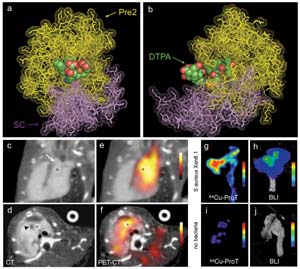 PET-CT image of S. aureus endocarditis.
The MGH Centre for Systems Biology, research team has explained the existence of a staphylococcus aureus-related endocarditis in a mouse prototype in their Nature Medicine Report, which was discovered using PET imaging along with a radio-labeled type of protein that normally hides the infectious micro-organisms like bacteria from the body immune system.
Endocarditis is an infection of the tissue surrounding the heart valve. The infection appears as formations known as vegetations that comprise clotting structures such as fibrins and platelets in combination with infectious micro-organisms. Diagnosing endocarditis is not easy, since the symptoms of endocarditis including heart murmur and fever, which are not very obvious and blood tests do not reveal s.aureus. Unless proper antibiotic therapy is taken, s.aureus endocarditis may completely destroy or harm the heart-valve.
The MGH research team initially examined the molecular mechanism due to which staphylocoagulase triggers the clotting process. The team found that one molecule of staphylocoagulase reacts with a minimum of four molecules of fibrin or fibrinogen, in a complicated pattern resulting in a growing vegetation. The team examined whether the labeled type of prothrombin could identify precisely s.aureus endocarditis in mice, as prothrombin is a vital intermediate product in the staphylocoagulase fibrin interaction.
Initial study showed that the optical imaging system, FMT-CT was able to identify a type of prothrombin that has a fluorescence label in the s.aureus-induced vegetation. The team then confirmed that with a radiolabeled typeof prothrombin , identification of s.aureus vegetation is possible, on combining FMT-CT and PET-CT imaging. This technique can be used in human-beings, subject to added advancements and FDA approval.