Towards the end of the 19th century, Wilhelm Conrad Röntgen was conducting experiments at the University of Würzburg on light phenomena and other emissions generated by a cathode tube known as Crooke’s tube. Crooke’s tubes consisted of glass bulbs with both positive and negative electrodes that exhibited a fluorescent glow when high-voltage currents were passed through the tube.

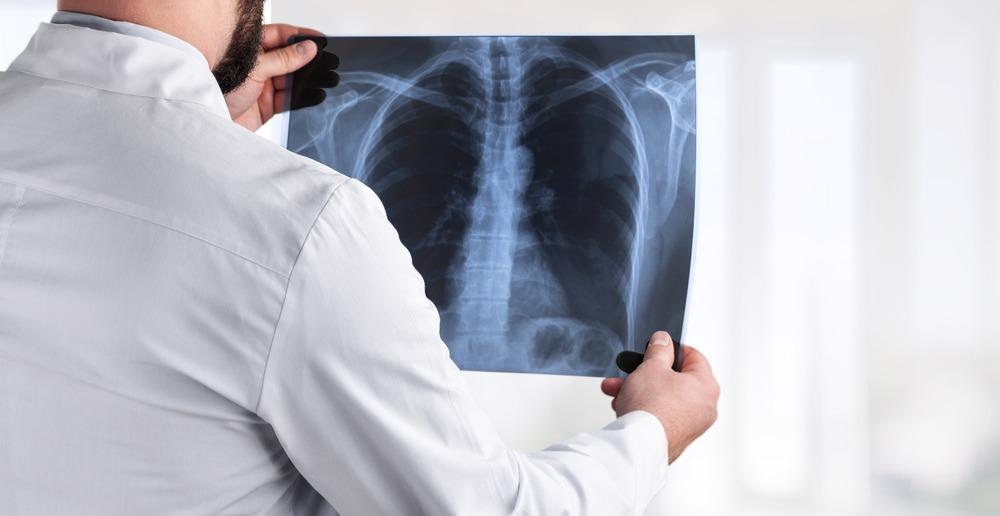
Image Credit: Billion Photos/Shutterstock.com
During this time, Röntgen observed the bones within his hand on a heavy black cardboard plate that was wrapped around the Crooke’s tube. From this observation, Röntgen deduced that the fluorescence he observed arose from invisible rays that were generated by the Crooke’s tube that were capable of penetrating the photographic plate that shielded the tube.
Further studies by Röntgen revealed that this new type of light ray was capable of penetrating through most substances, including soft tissues while allowing both bones and metals to remain visible. In addition to observing this phenomenon on his own hand, Röntgen famously imaged both the bones in his wife’s hand and her wedding ring in an image that has been regarded as the first X-ray.
Medical Imaging in the Early 20th Century
Soon after Röntgen published his findings in the Proceedings of the Würzburg Physico-Medical Society, researchers around the world were eager to apply his discovery to a wide range of applications. By February 1896, X-rays were used for the first time in a clinical setting in Dartmouth, Massachusetts, where Edwin Brant Frost used this technology to identify Colles fractures.
Despite the utility of this new approach to assessing skeletal trauma, as well as other health issues ranging from tuberculosis and other lung lesions to gangrene, many of the early medical imaging devices used extremely high doses of radiation to acquire these images. For example, film cassettes required patients to stand still for up to eleven minutes of intense radiation exposure during the medical imaging process.
By the 1940s, scientists began to understand the long-term risks associated with radiation exposure, which allowed for several changes to be made in how this imaging technology was used in the clinic. For example, lead aprons, gloves, and protective eyewear, as well as thyroid shields were quickly implemented into radiation safety procedures.
One way in which scientists looked to minimize patient exposure to radiation during medical imaging was using fluorescent screens in 1946. These screens were treating with fluorescence, while the treating clinician wore specialized glasses during the imaging process that allowed for them to identify anatomical structures. Despite the reduced risk of radiation exposure this new technique had on the patient, the doctor instead was at a greater risk of radiation exposure with this approach.
From the Mid-20th Century to Today
By the 1950s and early 1960s, the integration of information technology with X-ray technology arrived, revolutionizing medical imaging. Soon after in 1972, computed tomography (CT) was invented by British engineer Godfrey Hounsfield and South African physicians Allan Cormack.
The invention of CT imaging was based on the mathematical theory of Johann Radon in 1917 known as “Radon transform” combined with the “Algebraic Reconstruction Technique” described by Polish mathematician Stefan Kaczmarz in 1937.
By 1973, the first CT scan was used to image the brain of a woman who was believed to be suffering from a brain tumor. Interestingly, the entire imaging process took several days to complete, as the scanner took several hours to obtain the data needed to obtain an image of a single “slice” of the brain.
Although the invention of CT imaging allowed clinicians to gain much greater anatomical differentiation of body tissues, it also increased the dose of radiation that was administered to the patient as well.
A CT chest exam in 1991 averaged a radiation dose of 8 milliGray (mGy), which was about 200 times greater than the radiation dose administered in an ordinary X-ray exam at the time.
Since its original discovery, several advanced CT technologies have been developed to minimize the potential risks of exposing patients, particularly pediatric patients who are more vulnerable, to the effects of ionizing radiation exposure.
Some of the most notable CT techniques that are associated with a decreased scan time and radiation dose include dual-energy CT (DECT), imaging with a low tube potential, and rapid scanning.
During the 1970s, several years of investigation into magnetic resonance imaging (MRI) devices also led to the first MRI image of the brain in 1978.
The successful development and clinical application of this imaging technology were particularly important in the field of neuroscience, where researchers were able to advance their knowledge and understanding of various neuroanatomical regions. In doing so, researchers began to understand human memory systems, as well as associated neurodegenerative pathophysiological mechanisms.
Although MRI was more recently discovered, this medical imaging approach has also been expanded into both structural and functional MRIs.
Although MRIs are more time-consuming and require an enclosed environment to obtain the image, they avoid the use of ionizing radiation that is associated with X-ray imaging techniques.
References and Further Reading
Scatliff, J. H., & Morris, P. J. (2014). From Röntgen to Magnetic Resonance Imaging. North Carolina Medical Journal 75(2); 111-113. https://ncmedicaljournal.com/
November 8, 1895: Roentgen’s Discovery of X-Rays [Online]. Available from: https://www.aps.org/publications/apsnews/200111/history.cfm.
A Brief History of Medical Diagnostic Imaging [Online]. Available from: https://www.pbmchealth.org/news-events/blog/brief-history-medical-diagnostic-imaging.
Abdallah, Y, M. (2017). History of Medical Imaging. Archives of Medicine and Health Sciences 5; 275-278. https://www.amhsjournal.org/
Hessenbruch, A. (2002). A brief history of X-rays. Endeavour 26(4); 137-141. doi:10.1016/S0160-9327(02)01465-5.
History of CT Scan [Online]. Available from: https://catalinaimaging.com/history-ct-scan/.
Gottumukkala, R. V., Kalra, M. K., Tabari, A., et al. (2019). Advanced CT Techniques for Decreasing Radiation, Reducing Sedation Requirements, and Optimizing Image Quality in Children. RadioGraphics. doi:10.1148/2019180082.
Disclaimer: The views expressed here are those of the author expressed in their private capacity and do not necessarily represent the views of AZoM.com Limited T/A AZoNetwork the owner and operator of this website. This disclaimer forms part of the Terms and conditions of use of this website.