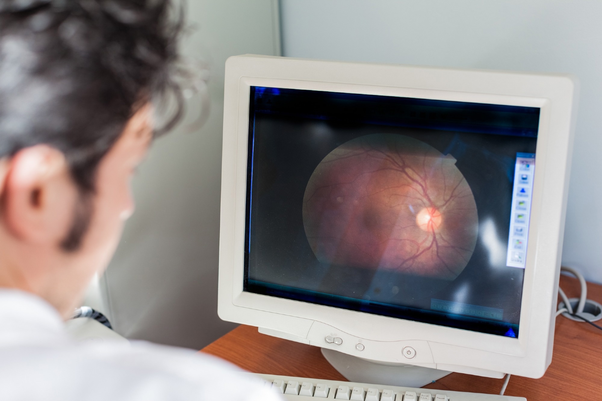
A recent article published in Scientific Reports explored the morphology of human cone photoreceptor mosaic. Researchers introduced a novel, fully automated algorithm to estimate cone spacing and density in adaptive optics (AO) montages. They also provided a publicly available database of normative images and cone densities.

Image Credit: Dario Lo Presti/Shutterstock
The goal was to enhance the understanding of cone density variations across the retina, which is crucial for improving clinical practices and research in ophthalmology.
Adaptive Optics Ophthalmoscopy
AO technology has transformed retinal imaging by enabling noninvasive observation of subcellular structures in healthy and diseased eyes. For over 25 years, AO ophthalmoscopy has allowed the correction of optical aberrations, achieving high-resolution imaging comparable to diffraction-limited systems.
By using wavefront sensors and deformable mirrors, this system compensates for ocular aberrations in real time, significantly enhancing image clarity. This advancement allows for more detailed studies of cellular integrity, particularly in retinal diseases, by revealing the structure and function of individual photoreceptors.
Despite its benefits, the wider clinical application of AO imaging has faced challenges, primarily due to the lack of automated tools for quantifying features in AO montages, such as the photoreceptor mosaic. Existing techniques often rely on manually selecting regions of interest (ROIs) and semi-automated algorithms for cone identification, which can introduce bias. Thus, there is a need for fully automated techniques to improve the accuracy and efficiency of AO imaging.
Algorithm and Dataset Development
The authors addressed these limitations by developing an automated algorithm that estimates local cone spacing and density across complete AO montages without manual intervention. They also created an open-access normative database of photoreceptor images, enhancing the usability of AO imaging by ensuring consistent and unbiased measurements of retinal structures.
The study included 50 participants with normal vision, aged 18 to 67, all without known retinal or corneal issues (with visual acuities of 20/25 or better). A multi-modal adaptive optics scanning light ophthalmoscope (AOSLO) captured high-resolution images of their retinas.
During image acquisition, participants were instructed to fixate on a target while their retinas were imaged, covering about 6 mm centered on the fovea. Both confocal and non-confocal methods were employed, and the images were processed using a custom MATLAB algorithm. This algorithm, operating in the Fourier domain, extracted cone spacing and density from overlapping ROIs, providing a detailed analysis of photoreceptor structures.
The researchers also compiled an open-source dataset, including cone density analyses based on retinal eccentricity across all four retinal meridians. They validated their results by comparing them with previous histological data, confirming that their automated approach was consistent with earlier findings.
Key Findings and Insights
The automated algorithm estimated cone densities across the dataset, with results aligning with prior studies. The average peak cone density (PCD) was 152,906 ± 53,209 cones/mm², consistent with known variations in cone density in the human retina. Densities were generally higher along the horizontal meridians than the vertical ones.
Significant variability in cone density was observed between participants, particularly near the fovea, where differences could reach up to 2.5 times. This variability decreased with distance from the fovea, dropping to 1.75 times at 1 mm away.
The study emphasized the importance of a normative database to account for individual differences in retinal structure. Additionally, total cone counts correlated with minimum cone density, showing that higher total counts did not always indicate higher densities.
The authors found that cone density was consistently higher along the horizontal meridians (temporal and nasal) than the vertical meridians (superior and inferior), with the standard deviation of cone density following a similar pattern. The automated approach reduced the need for manual input, improving efficiency and reliability in retinal imaging analysis.
Applications
This research has significant implications for clinical settings. The automated algorithm and database can assist ophthalmologists and scientists in assessing retinal health, especially for diagnosing and monitoring retinal diseases. By providing a standardized reference for cone density, the study improves diagnostic accuracy and treatment planning for conditions such as age-related macular degeneration and retinitis pigmentosa.
The open-access dataset also encourages collaboration and innovation within the ophthalmic research community, promoting the development of new methods and technologies for enhancing retinal imaging and analysis. The novel algorithm reduces the time and expertise needed for cone density assessments, making advanced retinal imaging more accessible in clinical practice.
Conclusion and Future Outlooks
The novel dataset and automated algorithm proved effective in advancing the understanding of the human cone photoreceptor mosaic. The authors highlighted the importance of standardizing cone density measurements and provided insights into variability in retinal structures. Their work aims to improve the clinical utility of AO imaging and pave the way for future research into retinal health and disease.
Journal Reference
Cooper, RF., et al. (2024). Morphology of the normative human cone photoreceptor mosaic and a publicly available adaptive optics montage repository. Sci Rep. DOI: 10.1038/s41598-024-74274-y, https://www.nature.com/articles/s41598-024-74274-y
Disclaimer: The views expressed here are those of the author expressed in their private capacity and do not necessarily represent the views of AZoM.com Limited T/A AZoNetwork the owner and operator of this website. This disclaimer forms part of the Terms and conditions of use of this website.