Reviewed by Lexie CornerMay 6 2024
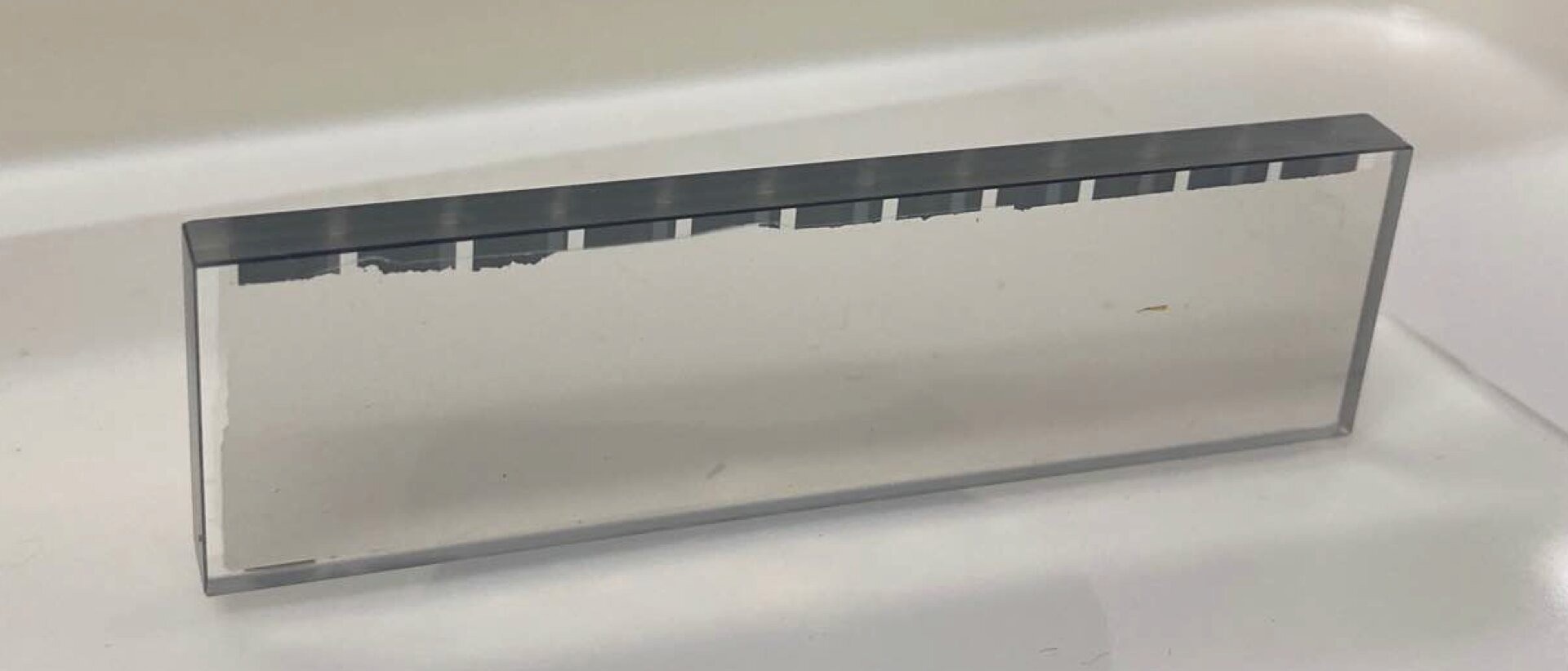
A Japanese group of scientists has created an X-Ray mirror with a pliable shape, resulting in exceptional atomic precision and enhanced stability. The research is published in the journal Optica.
 X-Ray microscopic images showing the higher resolution using the new deformable mirror. The left and right were obtained before and after shape correction, respectively. Image Credit: Matsuyama lab, Nagoya University
The latest technology, spearheaded by Satoshi Matsuyama and Takato Inoue at Nagoya University's Graduate School of Engineering, in partnership with RIKEN and JTEC Corporation, enhances the capabilities of X-Ray microscopes and other X-Ray mirror-based technologies.
An X-Ray microscope is an advanced imaging instrument that effectively bridges the gap between electron and light microscopy techniques. It uses X-Rays, which offer higher resolution than light and penetrate samples that are too thick of electrons. This makes it possible to image structures that are hard to view using conventional microscopy methods.
Due to their excellent resolution, X-Ray microscopes are particularly useful in disciplines like biology and materials science, where they may be used to observe the structure, composition, and chemical state of samples.
Mirrors are essential components of X-Ray microscopes. They provide high-resolution imaging of intricate objects by reflecting X-Rays. Exact measurements and high-quality photographs are essential, especially in cutting-edge scientific domains like catalyst and battery checks.
X-Rays are susceptible to distortion from minute production defects and external factors due to their short wavelength. Wavefront aberrations are produced, which may reduce the resolution of the image. Matsuyama and his associates resolved this issue by developing a mirror that can deform and change its shape in response to an X-Ray wavefront that is detected.
The researchers investigated piezoelectric materials to optimize their reflection. The ability of certain materials to deform or change shape in response to an applied electric field makes them valuable. As a result, the material may adapt to even small distortions in the detected wave by reshaping itself.
Upon evaluating multiple substances, the scientists ultimately selected a solitary crystal of lithium niobate to serve as their metamorphic mirror. Single-crystal lithium niobate can be polished to a highly reflective surface and extended and contracted by an electric field, making it valuable in X-Ray technology. This simplifies the device by enabling it to function as both the actuator and the reflecting surface.
Conventional X-Ray deformable mirrors are made by bonding a glass substrate and a PZT plate. However, joining dissimilar materials is not ideal and results in instability. To overcome this problem, we employed a single crystal piezo material, offering exceptional stability because it is made of a uniform material without bonding. Due to this simple structure, the mirror can be freely deformed with atomic precision. Moreover, this precision was maintained for seven hours, confirming its extremely high stability.
Satoshi Matsuyama, Department of Materials Physics, Graduate School of Engineering, Nagoya University
Matsuyama's team discovered that their X-Ray microscope performed better than expected when tested; due to its great resolution, it is particularly well suited for examining microscopic objects, including the components of semiconductor devices.
Their technique offers the potential to produce a microscope with a resolution around ten times greater (10 nm) than that of conventional X-Ray microscopy, which is usually only about 100 nm. This is because the aberration correction puts the resolution closer to the ideal value.
This achievement will promote the development of high-resolution X-Ray microscopes, which had been limited by the precision of the fabrication process; these mirrors can be applied to other X-Ray apparatus, such as lithography devices, telescopes, CT in medical diagnostics, and X-ray nanobeam formation.
Satoshi Matsuyama, Department of Materials Physics, Graduate School of Engineering, Nagoya University
Journal Reference:
Inoue, T., et al. (2024) Monolithic deformable mirror based on lithium niobate single crystal for high-resolution X-ray adaptive microscopy. Optica. doi.org/10.1364/optica.516909.