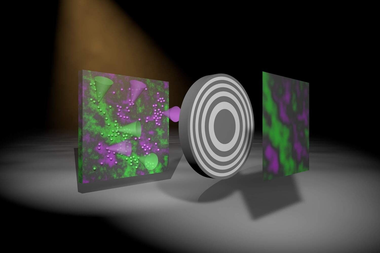
A research group has developed a new method to produce X-Ray images in color at the University of Göttingen.
 Artistic representation showing how an image is created using the newly developed method. Two colors–green and magenta–are emitted by fluorescing atoms in the sample (left) due to X-Ray excitation. The grey round object represents an optic casting a shadow on the detector. The algorithm then produces an actual image with two colors–the intensity of which represents the density of the fluorescing atoms within the sample. Image Credit: Markus Osterhoff.
Earlier, the only way to identify the sample’s chemical composition and the position of its components using X-Ray fluorescence analysis was to concentrate on the X-Rays and scan the entire sample. However, this process is costly and consumes a lot of time.
Researchers have currently come up with a method enabling an image of a large area to be generated from a single exposure without having to scan or focus. The method was reported in the Optica journal.
Contrary to visible light, there are no similarly strong lenses for “invisible” radiation, like X-Ray, gamma, or neutron radiation. But such kinds of radiation are necessary, for instance, in radiology and nuclear medicine, as well as in material analysis and industrial testing.
Uses for X-Ray fluorescence involve examining the composition of chemicals present in paintings and cultural artifacts to identify origin, authenticity, and production methods, or the analysis of soil samples or plants for environmental safety.
Also, the quality and purity of computer chips and semiconductor components can be checked through X-Ray fluorescence analysis.
For their newly-developed technique, the researchers made use of an X-Ray color camera developed by PNSensor in Munich and a novel imaging system that basically comprises a specially structured, gold-coated plate between the object and the detector, implying the sample casts a shadow.
In the detector, the intensity pattern quantified offers data regarding the distribution of the fluorescing atoms in the sample, which can further be decoded with the help of a computer algorithm. This new method implies that the plate could be close to the detector or object, unlike when using an X-Ray lens, which makes this a practical technique.
We have developed an algorithm that allows us to quickly and robustly create a sharp image, simultaneously for each X-ray color.
Dr. Jakob Soltau, Study First Author and Postdoctoral Researcher, Institute for X-Ray Physics, University of Göttingen
Co-author Paul Meyer, a doctoral student at the same institute, adds: “The optics simply cannot be compared to normal lenses; they were manufactured according to our precise specifications by a new company in Switzerland.”
This start-up company, named XRNanotech, specialized in nanostructures and was set up by Dr. Florian Döhring, who completed his Ph.D. at Göttingen University.
Next, we want to extend this approach to three-dimensional imaging of biological samples, as well as exploring phenomena in imaging such as inelastic scattering of X-rays, neutrons, or gamma radiation in nuclear medicine.
Tim Salditt, Professor and Research Group Leader, University of Göttingen
Journal Reference:
Soltau, J., et al. (2022) Full-field x-ray fluorescence imaging using a Fresnel zone plate coded aperture. Optica. doi.org/10.1364/OPTICA.477809.