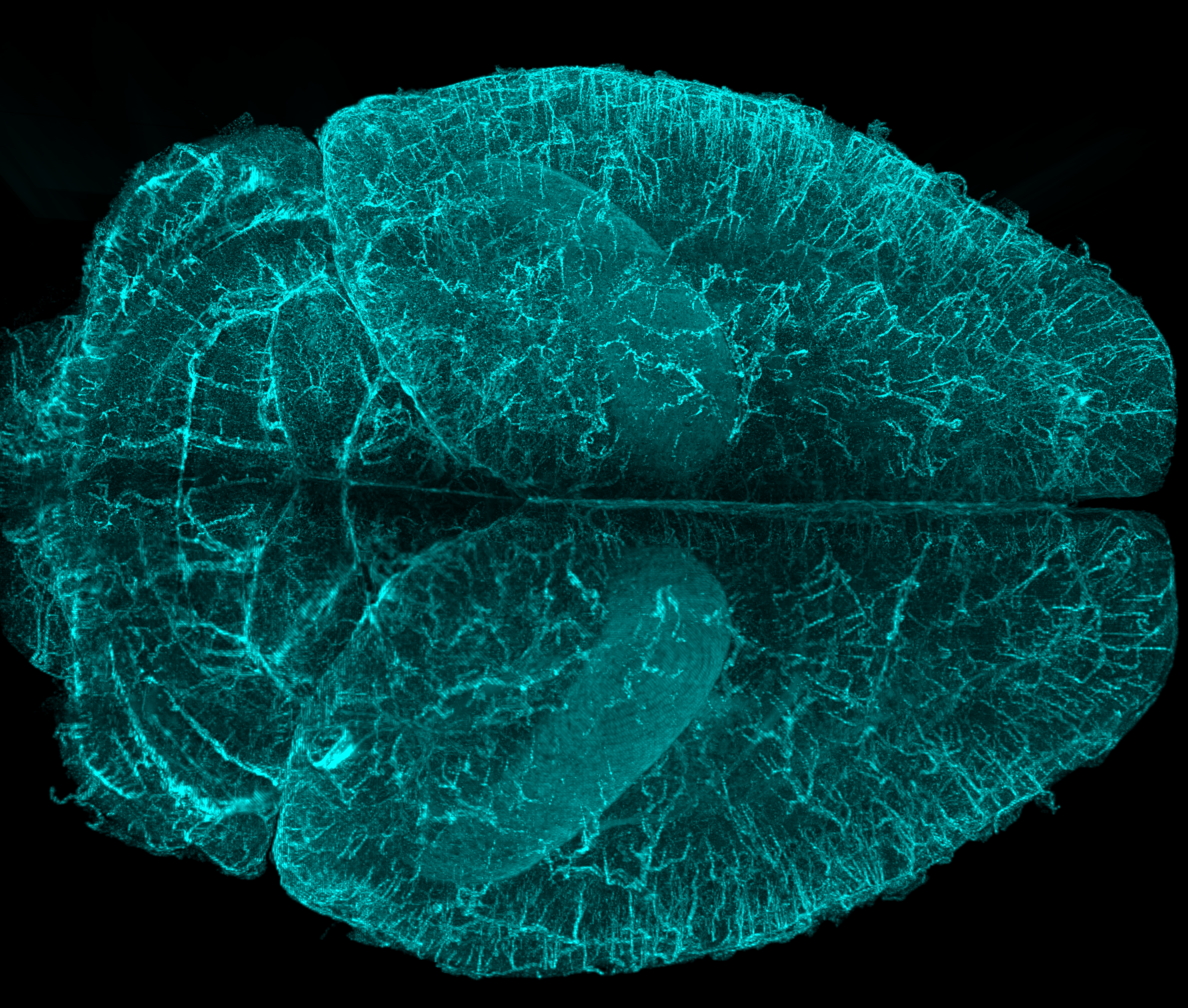
Image credit: Andor Technology Ltd.
Imaris, an Oxford Instruments brand, has today released Imaris 9.8, the latest version of its market-leading microscopy image analysis software.
In the latest release, we added object visualization on extended sections, together with the raw data which opens new ways of validating detection and editing in Imaris. Now, users can clearly see the precision of the Spots and Surfaces detection, even in a “cluttered” data volume with thousands of objects. These features build upon the previous improvements for the most widely used image analysis models, Spots and Surfaces, for big data analysis and smooth interaction.
Anyone who has ever traced neurons within datasets where dendrites are overlapping and occluding one another in the 3D space knows that tracing validation is an almost impossible task in the 3D view. In Imaris 9.8, neurons can also be presented together with the raw data on the 2D slice view. In addition, the section thickness and orientation can be freely adjusted in any direction in 3D, including oblique orientations.
This is not the only benefit Imaris 9.8 offers for neuroscientists, as the well-known Filament Tracer model is enriched with soma detection and modeling, which adds important cell body statistics and branching classification.
All Imaris and Imaris Viewer users will quickly discover that virtual dissections of datasets and the use of slicers are now much smoother and extremely intuitive. All clipping planes are translated and rotated using a new manipulator, easily operated with a mouse and keyboard.
“Having the ability to quickly inspect not only your images, but the analysis results from them is fundamental to image analysis,” said Meredith Price, Imaris Business Manager.
“The release of Imaris 9.8 builds upon our world-class rendering tools to simplify the validation and editing of analysis results furthering our commitment to intuitive usability for our customers.”
Imaris’ goal is to bring researchers the best and most complete user experience in the microscopy image analysis world. Users can download a 10-day free trial of Imaris to explore the software first-hand.