Mechanics play a pivotal role in the development, progression, and metastasis of cancer. Unfortunately, an effective and reliable method of mechanical characterization of metastatic cancer has remained elusive. Until this point, available methods had destroyed the tissue sample while analyzing it. In a new study published on the bioRxiv* preprint server, a team at the University of Sheffield, UK, present the findings of its latest research prior to peer review that demonstrate the efficacy of a novel method of characterizing metastatic breast tumors with atomic force microscopy (AFM).

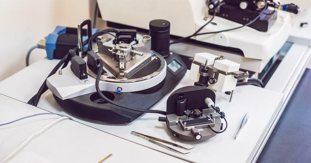
Image Credit: Elizaveta Galitckaia/Shutterstock.com
Paradoxical Evidence Regarding the Mechanical Properties of Breast Cancer
A wide body of research supports the notion that mechanics are fundamental to the development, progression, and metastasis of cancer. To better understand cancer, scientists have established two main methods of investigating cancer mechanics; those that measure the micro-properties of single cultured cells, and those that measure the properties of entire tumor tissues. However, evidence collected from these two methodologies has been contradictory. Data from studies on single cultured cancer cells have consistently demonstrated that these cells are more compliant than healthy cells, whereas data obtained from studying the bulk properties of whole tumors have shown them to be stiffer than the surrounding tissue. Scientists have theorized that tumors exhibit stiff components in the peripheral stroma, which would explain the paradoxical findings outlined above.
Previous studies have reported that cancer progression in breast cancer is associated with a softening of epithelial tumor cells, whereas other studies have reported that breast tumorigenesis is characterized by extracellular matrix (ECM) remodeling and stiffening. These contradictory findings demonstrate the heterogeneity of the mechanical properties of the tumor microenvironment. There is a need to deepen our understanding of the mechanics that underlie the later stages of cancer metastasis. Currently, few published studies exist on this topic and a reliable method of mechanical characterization is yet to be established.
To address this knowledge gap and the need for an effective characterization tool, scientists at the University of Sheffield conducted a study that tested a novel, AFM-based method of quantifying the mechanical properties of metastatic breast tumors.
An AFM-Based Method of Characterizing Metastatic Cancer
The team based in the UK developed a method to quantify the micro-mechanical properties of samples of metastatic breast tumors that remained relatively intact, as well as their surrounding bone microenvironment, utilizing atomic force microscopy. Its novel method quantified the micro-mechanical properties of breast cancer, including the elastic moduli and viscosity, without destroying the samples of breast cancer bone metastases and the surrounding microenvironment.
A mouse model was used to test the AFM-based approach. A commonly used in vivo mouse model was used, in which breast cancer bone metastases were established in mice via intracardiac injection of MDA-MB-231luc/GFP breast cancer cells. The team gathered details of the mechanical profile of the metastatic tumors by applying point force (F) vs. indentation (δ) and creep measurements by AFM.
The methodology was successful at quantifying the micro-mechanical properties of the tumor and its surrounding micro-environment. The results were compared with those obtained from explanted subcutaneous breast tumors grown via murine models and single 2D-cultured breast cancer cells in-vitro. This comparison demonstrated the significant differences in the measured mechanical properties of 2D in vitro models and 3D samples (e.g. non-orthotopic tumors and metastatic tumors). The results also revealed that using the AFM model, no impact of the metastatic tumor was found on the surrounding environment at the mesoscale (>200 µm).
The team’s novel method of mechanical characterization of metastatic cancer was proven to be effective and will likely be fundamental to studying complex cancerous tissue and uncovering important information on the mechanical properties of cancer metastases in bone.
*Important notice
bioRxiv publishes preliminary scientific reports that are not peer-reviewed and, therefore, should not be regarded as conclusive, guide clinical practice/health-related behavior, or treated as established information.
References and Further Reading
Chen, X., Hughes, R., Mullin, N., Hawkins, R., Holen, I., Brown, N. and Hobbs, J., 2021. Atomic force microscopy reveals the mechanical properties of breast cancer bone metastases. https://www.biorxiv.org/content/10.1101/2021.06.01.446539v1.full
Levental, K., Yu, H., Kass, L., Lakins, J., Egeblad, M., Erler, J., Fong, S., Csiszar, K., Giaccia, A., Weninger, W., Yamauchi, M., Gasser, D. and Weaver, V., 2009. Matrix Crosslinking Forces Tumor Progression by Enhancing Integrin Signaling. Cell, 139(5), pp.891-906. https://www.sciencedirect.com/science/article/pii/S0092867409013531
Disclaimer: The views expressed here are those of the author expressed in their private capacity and do not necessarily represent the views of AZoM.com Limited T/A AZoNetwork the owner and operator of this website. This disclaimer forms part of the Terms and conditions of use of this website.