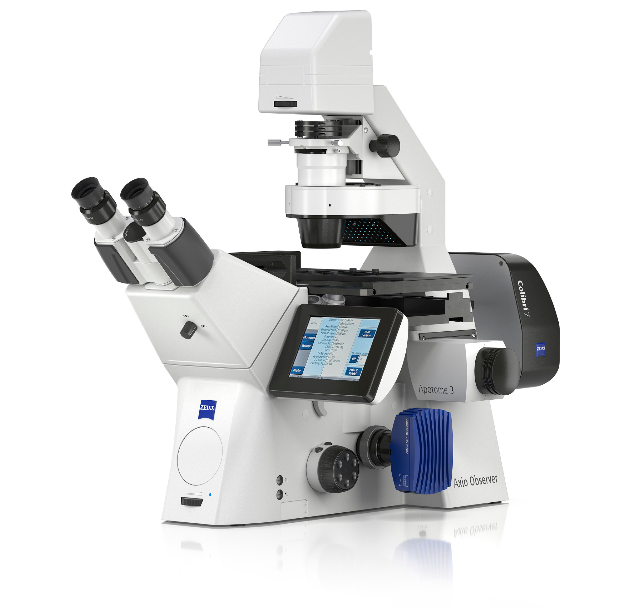
ZEISS presents the new AI Sample Finder for optimal user guidance and operation. With this feature, the open and flexible inverted microscope platform ZEISS Axio Observer makes sample placement easier than ever and significantly reduces the time to experiment.

For researchers, it offers a completely different way to operate a microscope, greatly boosting both productivity and ease of use. Users are guided more efficiently and have to perform fewer manual steps. They can navigate more intuitively and are able to analyze more samples on the carrier.
AI Sample Finder identifies the sample carrier and detects sample areas automatically. This accelerates the process of localizing a sample, especially when the sample is translucent or simply too small to see with the naked eye. After placing the sample on the loading position, AI Sample Finder moves it to the objective. There is no need to move microscope parts for sample placement manually. Focus is adjusted automatically, and a high-contrast overview image for fast and convenient navigation is taken within seconds even for very low-contrast samples. Intelligent routines reliably identify the sample carrier, regardless of its type. And with its deep learning algorithms, AI Sample Finder detects even unusual regions of interest.
In life science research, a good overview image is the foundation for a detailed analysis to make sure all areas can be navigated and accessed directly. AI Sample Finder eliminates time-consuming manual adjustments and reduces the time to image from minutes to just seconds. It also reduces phototoxicity because only short illumination of the sample is necessary when performing adjustments and identification of relevant areas. During the experiment, the entire system can be controlled remotely for highest flexibility.
New accessories for improved optical sectioning and efficient illumination
When imaging big specimens, out-of-focus light often blurs the image, thereby reducing contrast and resolution. With ZEISS Apotome 3, removing out-of-focus light becomes simple and efficient. It comes with three grids for structured illumination and creates crisp optical sections of fluorescent samples free of scattered light. Automatically recognizing the magnification, the ideal grid is moved into the beam path, Page 2 always resulting in the best image quality. The peer-reviewed algorithms yield artifact-free images even allowing the quantification of intensities. ZEISS Apotome 3 significantly increases the axial resolution compared to conventional fluorescence and adds optical sectioning capabilities to your widefield microscope for 3D rendering, even from thick specimens.
A new light source, the Excite Xylis LED is now integrated into the ZEISS microscopy environment. This effective illumination solution provides a high and reproducible light output over its entire life span without warmup times. With its broad emission spectrum, dyes from UV to far red can be excited for highest spectral flexibility. It is mercury-free and complies to latest RoHS standards. Long-lasting LEDs have resulted in changing light bulbs becoming obsolete and have led to lower running costs and positive outcomes for the environment.