Aug 7 2019
For the first time, scientists have integrated automated image analysis algorithms with a robust microscopy method to differentiate healthy tissues from metastatic cancerous ones without depending on the use of a contrast dye or invasive biopsies.
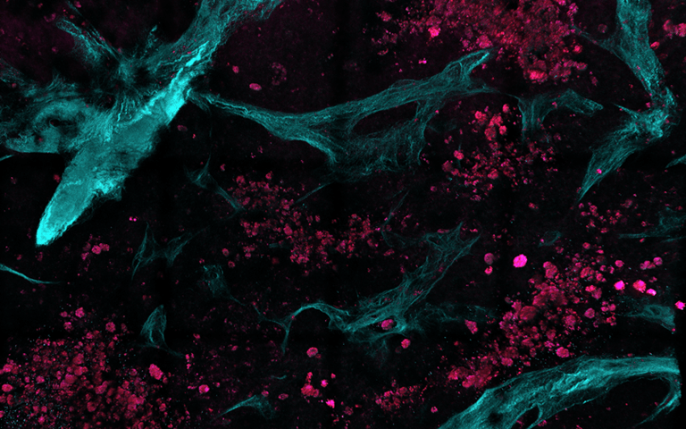
 The researchers combined multiphoton microscopy with automated image and statistical analysis algorithms to distinguish between healthy and diseased tissue. In this image, collected in a completely label-free, noninvasive manner, collagen is colored green while ovarian metastatic cell clusters are presented in red. (Image credit: Dimitra Pouli, Thomas Schnelldorfer, and Irene Georgakoudi, Tufts University and Lahey Hospital and Medical Center)
In the future, this latest technique may help physicians to detect cancer metastasis that is otherwise hard to visualize through normal imaging technologies at the time of operations.
“Existing techniques are invaluable but suffer from low spatial resolution and often require the use of exogenous contrast agents,” stated Thomas Schnelldorfer, research team co-leader from Lahey Hospital, Burlington, Massachusetts, U.S.A.
The method utilized in this work identifies in a completely label-free manner cellular and tissue features at the microscopic level, essentially acting like a biopsy without a knife.
Dimitra Pouli, Study Lead Author, Tufts University, Medford, Massachusetts, U.S.A
In Biomedical Optics Express, the Optical Society (OSA) journal, the researchers have described the application of multiphoton microscopy together with automated image and statistical analysis algorithms to inspect biopsies that were freshly removed from the peritoneal cavity—a part of the abdomen that is often impacted by metastatic cancers, particularly for patients suffering from ovarian cancer.
The study is the first to successfully assess healthy and metastatic human peritoneal tissue by integrating image texture analysis methods with microscopy modality.
Since the method assesses extracellular and cellular traits at the microscopic level, it can potentially detect cancer metastasis at an earlier stage when it might be easier to treat the condition. The approach uses algorithms to categorize tissues. In addition, it may help reduce bias in understanding images and complement techniques that depend on human expertise.
This could ultimately help surgeons identify suspicious or diseased areas directly in the operating room in real-time, which in turn would directly affect patient management.
Thomas Schnelldorfer, Research Team Co-Leader, Lahey Hospital, Burlington, Massachusetts, U.S.A.
“As the method exploits inherent tissue signals present almost ubiquitously in tissues, it can be applied to other types of cancer and other applications altogether, such as fibrosis and cardiovascular disease where tissue structure and extracellular matrix remodeling are altered by the underlying disease processes,” added Irene Georgakoudi, study co-leader from Tufts University.
Finding Clues in Tissue Texture
Multiphoton microscopy operates by transmitting laser light to tissues. Despite the high peak intensity of the laser, it is transmitted in extremely short pulses so that the average power is kept as small as possible and damage is not caused to the tissues.
When different tissue components communicate with the laser light, they produce signals that are subsequently retrieved by the microscope to generate an image. After acquiring the images, automated image processing algorithms can be utilized to expose special textural features. Such features, which cannot be seen in the images obtained with normal operative imaging tools, can now be examined with statistical models to categorize the tissue as diseased or healthy.
A major benefit of the technique is that the acquisition and analysis of images are predicated on the components of the tissue itself—like cells or collagen—and not on contrast dyes that have been introduced to it. Collagen is a protein that forms connective tissue. This approach makes it possible to analyze inherent features associated to form and function in a fully nondestructive and noninvasive way.
This is the first-ever study where this integrated microscopy and analysis technique was applied to healthy as well as metastatic human parietal peritoneal tissues. Since collagen is abundantly present in the parietal peritoneal tissue, part of the analytical implementation was concentrated on assessing the micro-structural patterns of collagen fibers as well as their intermolecular cross-linking signals.
Both healthy and diseased tissues were observed to have unique patterns in terms of correlation (a measure of pattern repetitiveness) and contrast (a measure of intensity differences from one pixel to another). While metastatic tissue images displayed smaller fibers and more uniform intensity patterns, healthy tissues displayed greater difference in these features.
Such changes demonstrate the negative impact of the cancer cells on the native connective tissue, offering a hallmark of cancer metastasis.
Improving Cancer Staging
Establishing the locations and extent of cancerous spread, called staging, is important for effective treatment of cancer. Tools like white light laparoscopy and cross-sectional radiographic imaging are used to detect abdominal metastases; however, these tools are not usually appropriate when it comes to identifying tinier lesions embedded inside the healthy tissue.
In addition, biopsies and microscopic assessment have an important role to play in establishing whether cancerous cells have metastasized and started to enter the tissue microenvironment.
When ovarian cancer starts spreading, it usually emerges first in the peritoneum—a kind of membrane that lines the abdominal cavity. The researchers tested their latest technique by examining peritoneal biopsies obtained from eight patients diagnosed with suspected or confirmed ovarian malignancy.
After examining 41 images obtained from the biopsies, the method precisely categorized 40 out of 41 images (with an accuracy of 97.5%). In total, 29 of 30 were accurately classified as healthy (96.6% specificity) and 11 samples were properly classified as metastatic (100% sensitivity).
Next, the scientists are planning to further test their technique in a larger sample of images acquired from a wider patient population. Although the analysis technique was designed for identifying ovarian cancer that has spread to the parietal peritoneal tissue, the same method can be adapted for examining other types of cancers and other types of tissues.
While the method was tested using biopsies, the end goal is to use it directly on the body areas where cancer is suspected or detected, without the necessity for dyes or biopsies, stated the researchers.
Before the method can be utilized for real-time tissue analysis at the time of surgery, more research will need to be done to combine the microscope with surgical instrumentation, reduce the size of the microscopy components, and allow real-time analysis of the obtained images directly at the operating room.