When seen under the microscope, subjects ranging from algae to zebrafish look attractive, strange and astonishing. Incredible views of the unseen universe won top prizes in the competition.
William Lemon, Philipp Keller and Fernando Amat belonging to the Howard Hughes Medical Institute Janelia Research Campus won the first prize for capturing an amazing video of the development of a fruit fly. The competition had received nearly 2,500 entries.
The winning movie captured a trembling ball of cells that turned into a fly larva in a fully developed state. By the end of the movie, the larva began to crawl off the screen. The first prize earned $5,000 worth of Olympus equipment, and the team is donating their prize to a new museum to be opened in 2015 in Northern Virginia named as the Children’s Science Center.
The other images that won awards are available for viewing on the Olympus BioScape website. This is the 11th year Olympus BioScapes Competition’s existence. This competition honors movies and images of plants, animals and humans that are captured using light microscopes. All images related to Life Science are eligible for taking part in the competition.
The criteria for judgement are the science represented, the impact, the attractiveness and the technical proficiency involved in capturing the images. Light microscopes of any brand may be used for capturing the images. This year, the competition honored the Top 10 award winners, and distributed one Technical Merit Award and 62 Honorable Mentions. Amongst these, nine movies won awards.

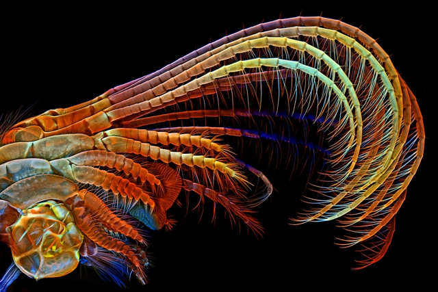
3rd Prize: Barnacle appendages. Image credit: Igor Siwanowicz / Olympus BioScapes
The most recent advances in developmental biology and neuroscience that have been documented by researchers have won prizes in this year’s competition. The First Prize movie is a high-speed video that captures what happens to individual embryonic cells from their fertilization stage to the period when the fly larva hatches.
During this period, division, migration, diversification and organ formation of the fly takes place. This movie enables researchers to understand the way in which an animal develops and the manner in which various cells are able to develop their unique functions. This development may help advance research into various diseases.
The team used a simultaneous multi-view light sheet microscope that was custom-built to capture the fruit fly movie that consisted of over two million images. A complex mathematical model helped extract the true potential of the data that allowed researchers to know what happened to each cell.
Thomas Deerinck, of the University of California San Diego’s National Center for Microscopy and Imaging Research, captured a dazzling photo of a rat cerebellum using multiphoton imaging at 300x. This photo won the Second Prize.
Igor Siwanowicz, a researcher from the HHMI Janelia Research Campus, captured a colorful confocal image of barnacle’s parts when they swept food into its shell for intake. This image earned the Third Prize. Csaba Pintér of Keszthely, Hungary had captured a funny image of two weevils, which won the Fourth prize.
This year’s competition saw participants from 68 nations. Images provided by participants from 21 nations and 14 states of the U.S. won honors. The other nations were Belgium, Chile, Canada, France, England, Germany, Denmark, Italy, Ireland, The Netherlands, Panama, Russian Federation, Spain, the United Arab Emirates, Sweden, Singapore, Poland, Norway, Japan, Iran and Hungary.
The list of other Top 10 prizewinners: Madelyn May, of Hano captured a confocal image of rat brain cerebral cortex which won the Fifth Prize; The Sixth Prize was for confocal image of a worm larva from plankton captured by N.H. David Johnston, of Southampton General Hospital, Southampton, UK; The Seventh Prize was won by butter daisy’s fluorescence photo which was imaged by Oleksandr Holovachov, of Ekuddsvagen, Sweden. A confocal image of the mouthparts of a vampire moth won the Eighth Prize and it was captured by Ashley L. Lash and Matthew S. Lehnert of Kent State University at Stark, North Canton, Ohio.
The confocal image of gears of a green cone-headed planthopper nymph captured by Igor Siwanowicz won the Ninth Prize. These planthoppers have the ability to accelerate at 500 times the force of gravity. In order to synchronize their hind leg movement, a pair of cogs and their trochanters are coupled. These images show that gears even exist in the natural world and are not necessarily a human invention.
Philipp Keller, Misha Ahrens and Fernando Amat of HHMI Janelia Research Campus had captured a video of an entire living zebrafish brain’s neural activity, and this entry won the Tenth Prize. Fast 3D recordings of the complete larval brain having about 100,000 neurons were captured by the video. This provides the most detailed image of single-neuron activity in a vertebrate’s brain.
For 11 years, Olympus has sponsored this competition to shed light on the importance of research and draw attention to the amazing intersection of science and art.
Olympus BioScapes movies and images have spurred public interest in and support of microscopy, drawn attention to the vital work that goes on in laboratories worldwide, and inspired young people to seek careers in science.
Hidenao Tsuchiya
Chairman of Olympus Scientific Solutions Americas
Part of the Olympus Corporation
In 2015, a museum tour is to travel around the U.S. and it displaying some of the Honorable Mention and winning images and videos. Olympus has sponsored the 2014 BioScapes tour in collaboration with Scientific American. Other winning BioScapes images are to be exhibited across Canada, Latin America, Mexico and the U.S.
The judges selected for BioScapes Digital Imaging Competition 2014 are eminent authorities in microscope imaging. The panel of judges selected by Olympus include Robert E. Campbell, Ph.D., University of Alberta, Edmonton, Alta., Canada; Alison North, Ph.D., The Rockefeller University, N.Y.; Catherine Galbraith, Ph.D., Oregon Health and Science University Knight Cancer Institute, Portland Ore.; and Douglas Murphy, Ph.D., HHMI Janelia Research Campus, Ashburn, Va.; Michael W. Davidson, The Florida State University, Tallahassee, Fla., directed the other judges in the competition.
The Olympus BioScapes Competition was founded to inspire brilliance in scientific photography and science. Enthusiasts are allowed to submit up to five movies of life science subjects, image sequences or still images that are captured using a compound light microscope. Brand and participant information is not provided to the judges.