Similar to the integration of supplemental optics in Hubble Space Telescope in 1990, an Italian research team has recently achieved a sight-correcting accomplishment for a microscope imaging method devised to study the universe appearing as huge as the Hubble Space Telescope but at the other end of the size spectrum , that is, the brain’s neural pathways.
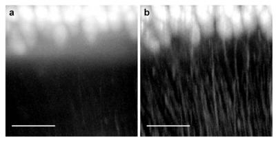
 This shows Purkinje cells from a mouse cerebellum imaged (a) with light-sheet microscopy and (b) with the significantly higher contrast provided by confocal light-sheet microscopy. The scale bar at the bottom is 100 micrometers across. Credit: Credit: Optics Express/European Laboratory for Non-Linear Spectroscopy, University of Florence, Italy
Light-sheet based microscopy (LSM), aka single plane illumination microscopy (SPIM), is an advanced microscope imaging technique, wherein laser-generated thin sheet of light illuminates a biological sample. However, radiance is directed from the side unlike other light sources. Fluorescence reflecting from the illuminated sample is directed upward via a lens, followed by being focused and captured using a digital camera.
Only one part of the target is imaged at a single given time, because the light sheet lights up the portion of the sample directly in the same plane. A series of 2-D sectional views called slices are generated rapidly by rotating the sample and by raising and lowering the illumination plane. By joining 2-D visual pieces, 3-D map of organs/systems or a whole organism can be produced.
Based on LSM, researchers analyzed the neural pathway in mice, a pathway vital for the functioning of the brain. High-resolution images of mouse brain tissues were obtained using the LSM method. The emitted light, scattered by whole brain samples, produces a background fluorescence that blurs the image, making it difficult to reconstruct the neural network.
To overcome this issue, Pavone and his co-workers integrated the features of light sheet illumination with confocal microscopy, in which a filter is used to remove photons scattered from the single plane of the thin sheet.
The background rejection capabilities of Confocal Light Sheet Microscopy (CLSM) were verified by demonstrating its potential to generate sharper images. While viewing three samples from mouse brains such as the cerebellum, the hippocampus, and the whole brain, CLSM was found to be more efficient than the other two methods. The 3-D reconstruction ability of CLSM was tested for mapping the micron-scale neuroanatomy of mouse Purkinje cells, which are huge neurons in the cerebellum, and locating an entire brain's neuronal projections.