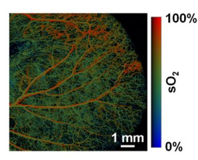
Photoacoustic tomography can penetrate several inches beneath the skin and capture images.
 The arteries (red) and veins (green) stand out clearly in a photoacoustic microscope image of a mouse ear. The new imaging technique, which marries sound and light to garner optical quality images of tissues beneath the skin is very sensitive to color changes like those that occur as hemoglobin becomes saturated with oxygen.
The advancements in photoacoustic imaging were presented by Lihong V. Wang, PhD, the Gene K. Beare Professor of Biomedical Engineering in the School of Engineering and Applied Science at St. Louis-based Washington University, in the scientific publication released March 23.
The photoacoustic tomography is one of the most striking advancements that demonstrate the utilization of oxygen by tissues. As hypermetabolism forms the basis of cancer, early detection will help reduce the associated risks.
The photoacoustic tomography is capable of converting the deeply- absorbed light to sound waves. A nanosecond-pulsed laser is used to irradiate the tissue to be imaged, at an optical wavelength. Light-absorption by molecules under the surface results in a thermally induced pressure that creates sound waves. These waves are measured by ultrasound receivers at the surface, organized to produce a photograph.
Light ensures a safe penetration. The photoacoustic images contain colored molecules that act as endogenous contrast agents, which comprise genes, hemoglobin, and melanin, and therefore show higher contrast compared to X-ray images.
The exogenous contrast agents like organic dyes or genes that can create colorful products enable photoacoustic tomography imaging of tissues including lymph nodes. Reporter genes also support photoacoustic images.
Enhancement in photoacoustic imaging is represented in Sentinel node biopsy, wherein a surgeon inserts a radioactive substance or a dye, close to a tumor. This is followed by dissecting the area and detecting the sentinel lymph node using the dye. However, this procedure has certain limitations. Wang’s new approach includes introducing an optical dye to obtain photoacoustic images wherein the sample can be extracted with a hollow needle.
Photoacoustic tomography is used to detect the oxygen saturation of hemoglobin and in various biological systems. Photoacoustic images also support the measurement of vessel cross-section and concentration of hemoglobin and blood flow speed.