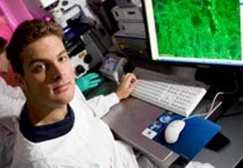
In recent times, image acquisition technology and microscopy have been modernized.
 The understanding of diseases such as Parkinson's and Alzheimer's is set to take a step forward following groundbreaking technology which will enable cell analysis using automated 3D microscopy.
The advanced microscopes can produce enormous data-sets in multiple dimensions which can occupy the space of the whole hard drive. These microscopes can eliminate manual error and reduce time consumption for processing these data-sets.
The Eskitis Institute for Cellular and Molecular Biology and the Griffith's School of Information Communication Technology have developed a novel technology to be used in automated three dimensional (3D) microscopes. The new technology enables automated examination, separation and detection of cells including neurons in the brain. Parkinson’s disease and Alzheimer’s disease can be understood more clearly by using this automated technology.
Griffith's Imaging and Image Analysis Facility Manager, Dr Adrian Meedeniya stated that many data-sets can be superimposed by using this automated technology in 3D microscopes and a thorough examination can be carried out by physicians and researchers by employing 3D microscopes instead of 2D microscopes.
Further, he added that the major inspiration to set up the partnership with the ICT School was to develop the automated technology, which can process large data-sets. The technology can be used to understand the effect of neuro-degenerative disorders on neurons of the brain, in a faster manner.
In future, the advanced technology with artificial cleverness can be broadly used for research in diseases.