Researchers at Rockefeller University have devised a new technique by controlling the specific attributes of polarized light. The method can be utilized to understand the orientation of particular proteins inside the cell. This can pave the way to study the functions of the nuclear pore complex.
According to Sandy Simon, who works as head of the Laboratory of Cellular Biophysics, the technique enables researchers to determine the arrangement of parts of huge protein complexes with respect to one another. It can also be utilized to investigate other multi-protein complexes of viral replication, protein synthesis and DNA transcription, he added.
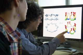
 A key component of the nuclear pore complex -- a Y-shaped cluster of proteins that helps determine what gets in and what stays out of a cell’s nucleus -- was first photographed and modeled at Rockefeller in 2009.
Sandy Simon and his research team comprising Martin Kampmann and Claire Atkinson have developed the new technique, which fills the ‘resolution gap’ between X-ray crystallography and electron microscopy, two techniques utilized to view protein complexes.
The researchers substituted the cell’s copy of the gene after linking the fluorescent markers genetically to separate parts of the nuclear pore complex. The replaced gene encodes the protein with the latest shape, which has the fluorescent tag. The researchers then measured the orientation of the light waves emitted by the fluorescently tagged proteins by utilizing special microscopes. By using both the latest measurements and information about the complex structure, the researchers can refute or validate the precision of previously recommended models.
The researchers utilized the technique to investigate nuclear pore complexes in human cells and budding yeast. In human cells, the numerous copies of the Y-shaped subcomplex are organized head-to-tail instead like the fence posts, validating a model recommended by Blobel in 2007.
According to the researchers, their new technique allows the measurement of proteins’ fluorescence while the cells are still active. Thus, the technique can be used to study the change in the structure of protein complexes over time and their reactions to different environmental conditions.