Optical imaging utilizes the unique properties of photons and light to obtain detailed images of cells, tissues, organs, and molecules. These techniques provide non-invasive or minimally invasive methods for examining the body.1

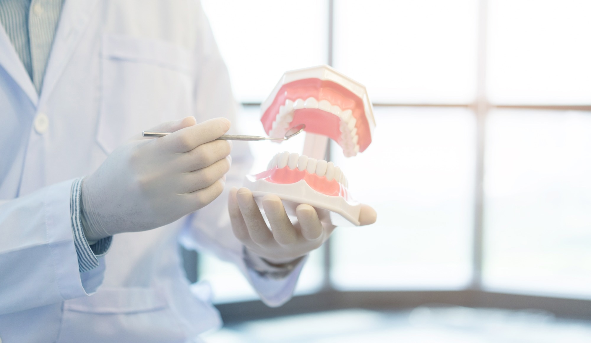
Image Credit: chainarong06/Shutterstock.com
Optical imaging significantly reduces the patient’s exposure to harmful radiation by using non-ionizing radiation, which includes ultraviolet, infrared, and visible light. Consequently, optical imaging can be used for repeated procedures to monitor disease progression or treatment outcomes, making it much safer than techniques requiring ionizing radiation, such as X-rays.1
In orthodontics, precise diagnosis and treatment plans are essential for excellent outcomes. Various optical imaging techniques are critical in enhancing diagnosis, treatment planning, and patient results. This article discusses the roles of intraoral scanning, optical coherence tomography (OCT), and scanning electron microscopy (SEM) in orthodontics.2
Optical Coherence Tomography
OCT is a modern imaging technique applicable to orthodontics. This tomographic imaging procedure has both optical reflection and absorption properties, allowing for the production of high-resolution cross-sectional images of teeth and soft tissues. Using the OCT technique, images of normal and pathological hard dental structures can be investigated, and the quality of different types of dental treatments can be assessed.3
This imaging method exhibits higher sensitivity and provides detailed imaging for early issue detection and treatment monitoring. For example, OCT can evaluate the periodontal ligament's responses to various orthodontic forces during treatment.3
Bracket debonding with fixed appliances is a crucial step at the end of orthodontic treatment. During this procedure, all adhesive resin from the enamel surface must be removed, and the tooth surface should be restored to its pre-treatment condition.3
Bracket debonding is a major cause of iatrogenic damage to enamel. OCT allows for minimally invasive visualization of near-surface alterations in complex tissues, providing real-time structural images of soft tissues and enamel through low-coherence interferometry using broadband light.3
A paper study published in Experimental and Therapeutic Medicine investigated the effects of bonding ceramic and metallic brackets on tooth enamel using OCT. Twenty permanent teeth were bonded and debonded using a side cutter/anterior bracket removal pliers.3
After the debonding procedure, the enamel, remaining adhesive, and bracket fragments on the tooth surface were investigated using OCT. Enamel cracks were confirmed only in samples bonded using ceramic brackets.3
Additionally, the type of pliers used did not influence the incidence or extent of enamel damage, nor did the debonding technique significantly affect the amount of adhesive remaining on the teeth. These findings suggest that the quality of materials and manufacturing processes for brackets can be enhanced by analyzing enamel structure using OCT.3
A recent study published in the Journal of Clinical Medicine verified that OCT can provide non-invasive three-dimensional (3D) and two-dimensional (2D) images of changes in hard and periodontal tissues during orthodontic treatment at the histological level in both animal experiments and humans in vivo.4
Thus, OCT devices can be effectively used in clinical practice, specifically for estimating microscopic changes in periodontal tissue during treatment. These changes are crucial for assessing the biomechanical effects and early periodontal disease diagnosis.4
Scanning Electron Microscopy
SEM analysis is an excellent approach for investigating the morphological and microscopic aspects of dental material surfaces. The technique can also be used to examine the surface microstructure of orthodontic mini-implants and evaluate cell adhesion.
SEM provides high-resolution images that enhance the understanding of interactions between biological tissues and implants.5 In orthodontic treatment, mini-screws optimize tooth movement by offering controlled anchorage. However, issues like partial osseointegration, device fractures, instability due to loosening, and soft tissue problems can occur.5
Typically, the choice of materials and design by the manufacturer influences how the implant interacts with surrounding tissues. In some cases, specific materials and designs can lead to unwanted responses in soft tissues and bone.5
A paper recently published in Scientific Reports investigated the impact of implant surfaces on cell growth and adhesion of human primary osteoblasts and fibroblasts using orthodontic TiAl6V4 mini-screws from three manufacturers. SEM was employed to qualitatively assess the cell-implant interaction at the drilling, neck, and top part of the screws.5
Although both cell types adhered to and grew on all three mini-screws, subtle differences were detected in cell shape and spreading based on the implant surface microstructure. This indicates that cell adhesion to implant surfaces can be controlled by manipulating machining conditions.5
In another recent study published in the Dentistry Journal, an SEM analysis of a polyetheretherketone (PEEK) retainer that had failed in an orthodontic patient was performed. After 15 months of use, the patient reported a gap opening between teeth 41 and 42.6
SEM was employed to evaluate the composition and microstructure of the retainer at various magnifications. The study findings suggested that the retainer's failure was multifaceted, implicating several factors such as patient-related issues, environmental influences, inadequate design, manufacturing flaws, and material defects.6
Thus, SEM analysis provided crucial insights into the mechanisms of PEEK retainer failure in orthodontic applications, which can inform the design and manufacturing of more effective PEEK retainers.6
Intraoral Scanning
An intraoral scanner is a device that integrates a software system, an intraoral camera, and a computer to capture detailed images of the oral cavity and teeth. Intraoral scanning offers real-time visual feedback for patients and clinicians, enhancing communication and understanding of treatment plans.7
Recent advances in intraoral scanning have improved the reproducibility and accuracy of recorded measurements, facilitating both treatment planning and diagnosis. A paper published in the International Journal of Environmental Research and Public Health reviewed the accuracy and reproducibility of intraoral scanners.8
The results showed that 3D models made using intraoral scanning methods provided clinically and statistically acceptable accuracy for all indirect and direct dental measurements. However, 3D models obtained using cone beam computed tomography (CBCT) underestimated dental measurements in the lower arch, although the measurements remained within clinically acceptable limits.8
Additionally, comparisons between the two intraoral scanners showed that the accuracy of the 3Shape TRIOS and the iTero Element intraoral scanners was clinically adequate. Both intraoral scanners exhibited excellent reliability and reproducibility. However, CBCT scanning and intraoral scanning of alginate impressions were valid and reproducible when reliability, validity, and reproducibility were examined collectively.8
Future Outlook and Conclusion
Optical imaging devices possess significant potential for diagnosing soft tissues, including hard and periodontal tissues, in the orthodontic field. In the future, OCT examinations may be utilized in vivo to aid orthodontic procedures in restoring tooth surfaces to their pre-treatment condition.3,4
Additionally, experience and strong clinical skills will be vital for the effective use of intraoral scanners.8 Companies, such as Launca and iTero, are some of the leading suppliers of optical imaging devices.
In summary, optical imaging techniques, such as OCT and intraoral scanning, could revolutionize orthodontics by providing more precise and accurate information about dental and periodontal tissues. As technology advances, these techniques are likely to play an even greater role in improving the quality of orthodontic care.
More from AZoOptics: Recent Developments in Brain Imaging Technology
References and Further Reading
- National Institute of Biomedical Imaging and Bioengineering. (n.d.). Optical Imaging. [Online] National Institutes of Health. Available at https://www.nibib.nih.gov/science-education/science-topics/optical-imaging (Accessed on 25 September 2024)
- Mohammed, KAK., Norehan, M. (2023). Accuracy of digital radiography in orthodontic diagnosis and treatment planning. World Journal of Current Medical and Pharmaceutical Research. https://wjcmpr.com/index.php/journal/article/view/282
- Petrescu, SMS. et al. (2021). Use of optical coherence tomography in orthodontics. Experimental and Therapeutic Medicine. DOI: 10.3892/etm.2021.10859, https://www.ingentaconnect.com/content/sp/etm/2021/00000022/00000006/art00028
- Baek, J.H. (2023). Potential Application of Non-Invasive Optical Imaging Methods in Orthodontic Diagnosis. Journal of Clinical Medicine. DOI: 10.3390/jcm13040966, https://www.mdpi.com/2077-0383/13/4/966
- Reimers, SN., Şen, S. (2024). Investigating adhesion of primary human gingival fibroblasts and osteoblasts to orthodontic mini-implants by scanning electron microscopy. Scientific Reports. DOI: 10.1038/s41598-024-68486-5, https://www.nature.com/articles/s41598-024-68486-5
- Zecca, PA. et al. (2024). Failed Orthodontic PEEK Retainer: A Scanning Electron Microscopy Analysis and a Possible Failure Mechanism in a Case Report. Dentistry Journal. DOI: 10.3390/dj12070223, https://www.mdpi.com/2304-6767/12/7/223
- Alshammery, FA. (2020). Three dimensional (3D) imaging techniques in orthodontics-An update. Journal of Family Medicine and Primary Care. DOI: 10.4103/jfmpc.jfmpc_64_20, https://journals.lww.com/jfmpc/fulltext/2020/09060/Three_dimensional__3D__imaging_techniques_in.7.aspx
- Christopoulou, I., Kaklamanos, EG., Makrygiannakis, MA., Bitsanis, I., Perlea, P., Tsolakis, AI. (2021). Intraoral Scanners in Orthodontics: A Critical Review. International Journal of Environmental Research and Public Health. DOI: 10.3390/ijerph19031407, https://www.mdpi.com/1660-4601/19/3/1407
Disclaimer: The views expressed here are those of the author expressed in their private capacity and do not necessarily represent the views of AZoM.com Limited T/A AZoNetwork the owner and operator of this website. This disclaimer forms part of the Terms and conditions of use of this website.