Verification of cancer diagnosis is done via histopathology by eliminating a part of the tissue in question after conducting imaging tests like CT, MRI, ultrasound or endoscopy.
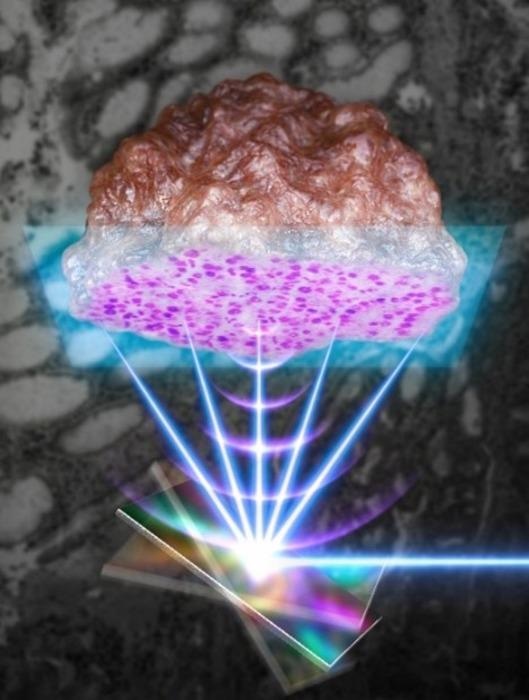
 Using a MEMS scanner that can scan the light and sound waves quickly, a high-speed pathological biopsy is performed on clinical tissue resected from cancer patients. Image Credit: Pohang University of Science and Technology.
Depending on the clinical diagnosis, the cancerous tissue is eliminated surgically and suspected tissues or lymph nodes are further analyzed. Additional treatment plans, like chemotherapy and radiation, are formulated on the basis of such outcomes. In recent times, a research group headed by POSTECH and Gachon University College of Medicine has come up with a machine learning-based histopathology technique.
Headed by Professor Chulhong Kim (POSTECH’s Department of Electrical Engineering, Department of IT Convergence Engineering, Department of Mechanical Engineering), the researchers collaborated with scientists from Gachon University College of Medicine to make a machine learning-based label-free histopathology device that exhibits the ability to execute histopathology in real-time.
Complex procedures, like freezing, staining or sectioning, are avoided by the device by making use of the ultraviolet (UV) photoacoustic imaging technology (UV-MEMS PAM) that intersects ultra-high-speed MEMS scanner technology. This study was chosen as the inside front cover paper in the September issue of Laser and Photonics Reviews, an international scientific journal in the field of optics.
At the time of cancer resection surgery, histopathologic examination is necessary to verify the site of the tumor. Traditionally, frozen-section testing is employed to perform the examination. However, as a result of its hard process, it can extend surgery and possibly produce interpretation errors.
To defeat these flaws, the research group suggested a high-speed reflection-mode UV photoacoustic microscopy system with the help of the 1-axis MEMS scanner as a novel label-free intraoperative histopathology technique.
With the help of this new method, the scientists were successful in observing label-free cell nuclei of mouse and human tissues. Additionally, by imaging clinical specimens that were resected from actual cancer patients and numerically measuring the histopathologic outcomes, the research team successfully illustrated that the suggested UV-PAM system has considerable potential as an alternative intraoperative histopathology method.
Photoacoustic imaging not only allows three-dimensional imaging without an autonomous contrast agent but also integrates the benefits of high-resolution optical imaging with ultrasound imaging that can realize structural and functional imaging of small cells and living tissues ranging to big organs. The microscope developed this time makes use of a high-speed MEMS scanner to considerably enhance the imaging speed.
The microscope developed in this study is new in that it distinguishes normal tissues from cancer tissues by performing photoacoustic histopathology on cancer tissues excised from actual cancer patients, and quantifying the pathological microstructures. This microscopy system is anticipated to dramatically cut down on the time it takes for biopsies during surgery, and to increase the stability and reliability of surgery and treatment.
Chulhong Kim, Professor, Pohang University of Science and Technology
This study was performed with the help of Korea’s Ministry of Science and ICT, the Ministry of Education, and the Ministry of Trade, Industry and Energy.
Journal Reference:
Baik, J. W., et al. (2021) Intraoperative Label-Free Photoacoustic Histopathology of Clinical Specimens. Laser & Photonics Reviews. doi.org/10.1002/lpor.202100124.