Jul 1 2021
According to a new study, thermal imaging techniques can determine whether a specific wound requires additional management, thus serving as an early alert system to enhance chronic wound care.
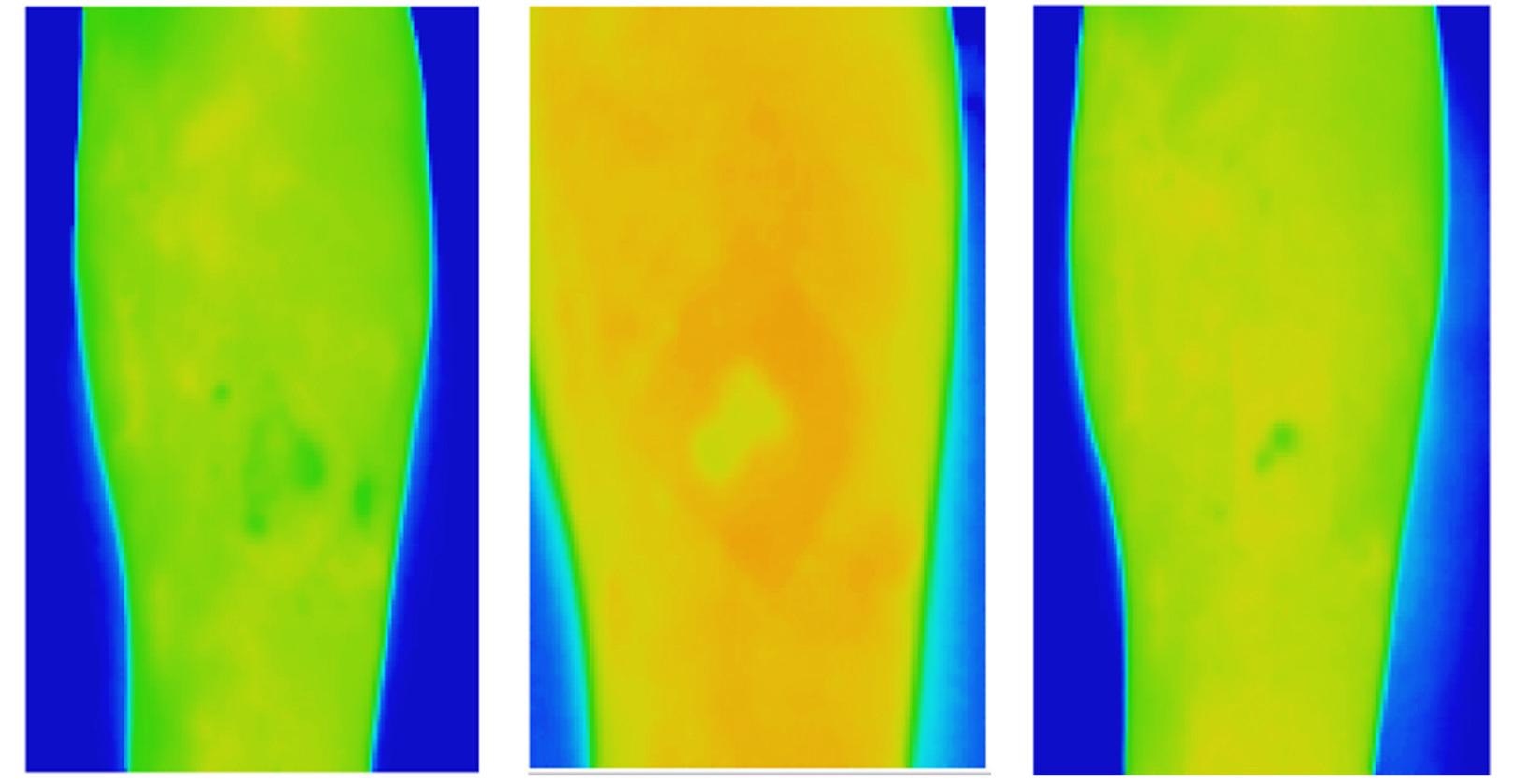
 Thermal images of a venous leg ulcer showing healthy healing progress over three weeks. Image Credit: RMIT University.
Chronic wounds affect almost 50% million Australians, costing the country’s health system approximately $3 billion per year. In fact, unattended chronic wounds are a major cause of limb amputation.
According to the Australian study, textural investigation of thermal images of venous leg ulcers (VLUs) can predict whether extra management needs to be devoted to a certain wound as early as the second week for people receiving treatment at their homes.
The clinical analysis performed by RMIT and Bolton Clarke is the first to explore the textural analysis on VLUs using thermal images that eliminate physical contact with the wound. The study was published in Scientific Reports, the Nature journal.
The team noted that the new technique, which provides data on spatial heat distribution in a wound, can precisely estimate whether the VLUs would heal in 12 weeks, by week two, after the baseline assessment.
The reason for this is a wound can change dramatically over the healing trajectory, with higher temperatures indicating potential infection or inflammation and lower temperatures indicating a slower healing rate because of reduced oxygen in the region.
According to Dr. Rajna Ogrin, a Senior Research Fellow from the Bolton Clarke Research Institute, the present gold standard for predicting the healing of VLUs — traditional digital planimetry — needs physical contact.
A non-contact method like thermal imaging would be ideal to use when managing wounds in the home setting to minimize physical contact and therefore reduce infection risk.
Dr Rajna Ogrin, Senior Research Fellow, Bolton Clarke Research Institute
After demonstrating that conventional thermal imaging techniques did not provide reliable results, the researchers devised a new technique for the analysis and applied this in the clinical trial.
The research work was carried out in 60 participants with VLUs and it revealed that thermal imaging provides an improvement over the present guidance for using planimetry wound tracings or digital imagery to detect the healing wounds by the fourth week.
The significance of this work is that there is now a method for detecting wounds that do not heal in the normal trajectory by week two using a non-contact, quick, objective, and simple method.
Dr Rajna Ogrin, Senior Research Fellow, Bolton Clarke Research Institute
According to Professor Dinesh Kumar from RMIT, standard wound photography cannot be easily used to accurately measure the differences in wound size and other physiological parameters over time in home-care settings.
This is because there are large variations between images due to changes in the lighting conditions, image quality, and differences in camera angle across specific points in time. Textural analysis of thermal images is resilient to these variations and is a time-efficient and cost-effective method to identify delayed healing of VLUs and improve patient outcomes.
Dinesh Kumar, Professor, RMIT University
Kumar also heads the Biosignals for Affordable Healthcare group in RMIT’s School of Engineering.
Journal Reference:
Monshipouri, M., et al. (2021) Thermal imaging potential and limitations to predict healing of venous leg ulcers. Scientific Reports. doi.org/10.1038/s41598-021-92828-2.