Nov 1 2018
Light-sheet fluorescence microscopy is a newly developed method that has given way to many biological findings by enabling scientists to produce 3D images of tissue—even live animal embryos—with the help of fluorescent tags.
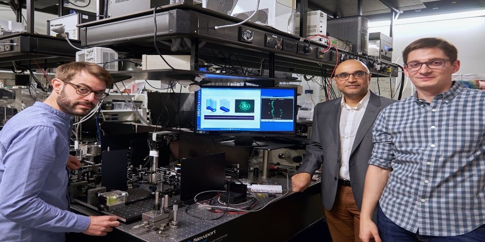
 For the first time, researchers have used three-photon absorption to increase the imaging depth of light-sheet fluorescence microscopy. Pictured are Federico Gasparoli, Kishan Dholakia, and Adrià Escobet-Montalbán with the newly developed three-photon light-sheet fluorescence microscope. (Image credit: Courtesy of the University of St. Andrews)
Now, with the use of an optical phenomenon called three-photon absorption, scientists report the potential to boost the imaging depth of light-sheet fluorescence microscopy.
In Optics Letters—The Optical Society (OSA) journal—the researchers have explicitly demonstrated how three-photon absorption can be utilized with light-sheet fluorescence microscopy for deeper imaging of tissues. As a demonstration, the team employed the combined method to create vivid images all through a ball of cultured cells, called a spheroid, which has a diameter of approximately 450 microns.
“This demonstration is very important as it addresses an unmet need of better imaging at depth, which could help scientists gain better data about biological processes,” stated research team leader, Kishan Dholakia of University of St. Andrews in the U.K. “This approach could be especially useful for neuroscience and developmental biology studies and might find application in imaging multiple samples in an automated way for drug discovery.”
Light is required for imaging fluorescent labels, but this light can be very damaging and even lethal to fragile biological samples, for example, animal embryos or brain tissue used for studying the development and disease processes. Light-sheet fluorescence microscopy enables rapid and high-resolution imaging without much optical damage, since it lights up a sample with only a thin sheet of light; unwanted light exposure is not experienced by other parts of the sample.
“We expect three-photon light-sheet fluorescence microscopy to make a large impact on imaging the brain in rodents such as mice and rats, where it could be used to capture very wide-field images with subcellular resolution,” said Adrià Escobet-Montalbán, the paper’s first author.
Deeper imaging
As part of the study, three-photon light-sheet fluorescence microscopy was compared to the formerly used two-photon absorption. The fluorescent label in multiphoton absorption emits light after absorbing, or being activated by, two or three photons instead of the one photon used for producing conventional fluorescence.
Multiphoton absorption not only minimizes out-of-focus light but also reduces light that can possibly damage the sample since it employs longer wavelengths, which are scattered relatively less by the tissue, and confines the excitation light to a small volume. These advantages are amplified when three photons are used for producing fluorescence instead of two.
The researchers demonstrated their novel method by using a typical optical setup for light-sheet fluorescence microscopy with a pulsed laser conventionally used for two-photon excitation. Even though this laser was not the most suitable for producing efficient three-photon excitation, it was perfect for comparing three-photon and two-photon excitation.
Next, the researchers used two-photon and three-photon excitation to image spheroids of human embryonic kidney cells. Close to the surface of the spheroid, both imaging modalities were seen to perform in a similar manner. Yet, at the distant side of the spheroid, the image quality of the two-photon image reduced significantly, while the image quality for the three-photon light-sheet fluorescence microscopy retained the image contrast.
Optimizing the technique
To additionally enhance the field of view and depth imaging, the team experimented with modifying the laser’s light intensity profile to a Bessel beam, which has a central bright core enclosed by concentric rings, instead of the standard solid Gaussian laser beam similar to that of a laser pointer.
“Bessel beams can be used in two-photon light-sheet mode but may yield potential artifacts due to their concentric rings,” said Federico Gasparoli, co-author. “For the first time, we show numerically and experimentally that these problems are suppressed in three-photon light-sheet fluorescence microscopy and that the beam goes even deeper, making this approach very attractive.”
Next, the team is planning to enhance the method by applying laser systems at longer wavelengths that are particularly developed for three-photon absorption. This should facilitate imaging at increased depth. At the same time, the researchers are also exploring to develop new ways to identify the light emitted from fluorescent labels deep within the samples.