Mar 24 2014
Using a University of Pennsylvania-designed device to noninvasively and continuously monitor cerebral blood flow (CBF) in acute stroke patients, researchers from Penn Medicine and the Department of Physics & Astronomy in Penn Arts and Sciences are now learning how head of bed (HOB) positioning affects blood flow reaching the brain.
Most patients admitted to the hospital with an acute stroke are kept flat for at least 24 hours in an effort to increase CBF in vulnerable brain regions surrounding the damaged tissue. Researchers report in the journal Stroke that, while flat HOB did indeed increase CBF in the damaged hemisphere in most stroke patients, about one quarter of the patients had a paradoxical response and showed the highest CBF with their head at an elevated angle.
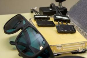
 The Penn-designed novel optical technique, called diffuse correlation spectroscopy (DCS), uses a noninvasive probe placed on the surface of the head to measure the fluctuations of near-infrared light caused by moving red blood cells in tissue, and has been shown to accurately track cerebral blood flow in underlying brain tissue. Credit: Rob Press for Penn Medicine
The Penn team has been developing and testing a new optical device that permits noninvasive and continuous monitoring of CBF at the patient's bedside. The key technology development is a noninvasive probe placed on the surface of the head that measures the fluctuations of near-infrared light that has travelled through the skull and into the brain, then back out to the tissue surface. These fluctuations are caused by moving red blood cells in tissue, and have been shown to accurately track CBF in underlying brain tissue. The novel optical technique, called diffuse correlation spectroscopy (DCS), proved to be more sensitive for detecting CBF changes with HOB positioning than the Transcranial Doppler (TCD), which uses acoustic waves to quantify blood flow velocity of the large arteries supplying the brain.
"This study illustrates the potential of using advanced technology to make individualized treatment decisions in real time" said senior author John A. Detre, MD, professor of Neurology and Radiology in the Perelman School of Medicine at the University of Pennsylvania. "While, on average, our findings support current guidelines to lay patients flat following stroke, they also suggest that for some stroke patients, lying flat may be either unnecessary or even harmful. Future studies examining clinical outcomes after stroke and using optical CBF measurements to guide management will be needed to confirm this."
Stroke is the leading cause of disability in industrialized nations and one of the leading causes of death, so even subtle improvements in stroke outcome can be expected to have a significant public heath impact. A reduction in CBF causes stroke, therefore most current interventions for stroke are intended to increase CBF. Yet, CBF is rarely measured in stroke patients, and if CBF is measured, it is usually a single measurement from a CT or MRI scan taken while the patient is lying flat. While CT and MRI are critical diagnostic tools used in stroke management, they are not well suited for assessing response to clinical interventions over time.
"We believe that these optical CBF measurements are detecting brain tissue blood flow of local microvasculature that might differ due to injury" states Arjun Yodh, PhD, a professor in the Department of Physics & Astronomy in Penn Arts and Sciences who has led the development of this new technology.
Among all patients, blood flow to the brain hemisphere where the stroke damaged tissue was reduced by 9 percent when the head of bed was elevated 15 degrees, and 17 percent lower when elevated 30 degrees. But in 29 percent of the patients, the optical method showed a "paradoxical" improvement in CBF when the bed was elevated. A prior study found almost the same proportion of "paradoxical" responders to HOB positioning, and in the combined cohort no clinical or radiological differences predicted an expected versus a paradoxical response.
"Our study suggests that it would be impossible for stroke clinicians to know whether HOB flat is optimal without actually measuring the response" said Michael Mullen, MD, a Penn stroke neurologist involved in the study. This may also be true for other clinical interventions such as administration of fluids, withholding antihypertensive therapies, or using medications to raise blood pressure. "The ability to measure cerebral blood flow continuously has tremendous potential and may one day allow clinicians to individualize therapy for each patient."
"We hope this technology will be able to guide management in advance of clinical symptoms" said Yodh. While the optical CBF instrumentation used for this study required a team of physicists to acquire and analyze the data, newer versions can be operated by clinical personnel and provide a real-time CBF display. Optical CBF monitoring may someday be performed routinely in all patients with acute brain injury.
The study team included Rickson C. Mesquita, PhD, now at the University of Campinas in Sao Paulo, Brazil, Christopher G. Favilla, MD, Michael Mullen, MD, Xiangping Lu, MD, Scott E. Kasner, MD, Joel H. Greenberg, PhD, and John A. Detre, MD from Penn's Department of Neurology; Meeri N. Kim, PhD, David L. Minkoff, and Arjun G. Yodh, PhD, from Penn's Department of Physics & Astronomy; and Turgut Durduran, PhD, now at the ICFO-Institut de Ciències Fotòniques, Castelldefels in Barcelona, Spain. Stroke patients were recruited from the Comprehensive Stroke Center at the Hospital of the University of Pennsylvania.
The study was funded by a Bioengineering Research Partnership grant from the National Institutes of Health's National Institute of Neurological Disorders and Stroke (NINDS - NS060653) as well as an NINDS Center Core in Neuroimaging (NINDS -NS058386), the Eunice Kennedy Shriver National Institute of Child Health and Human Development (NICHD - HD050836), and National Institute of Biomedical Imaging and Bioengineering (NIBIB - EB015893), along with the Sao Paolo Research Foundation and Fundacio Cellex.
Patent US#8,082,015 has been granted to the University of Pennsylvania for the optical CBF technology. Besides application to neurological disorders, it is being tested in a number of other clinical populations in whom CBF changes are relevant.