Feb 28 2014
Seventh- and eighth-grade students attending the March 10 “Brainworks” at Cedars-Sinai will see how a new, experimental device makes brain tumors glow – using a special camera, a laser, an imaging agent called Tumor Paint BLZ-100 and a synthetic version of a small protein found in the venom of the deathstalker scorpion.
 Seventh- and eighth-grade students attending the March 10 “Brainworks” at Cedars-Sinai will see how a new, experimental device makes brain tumors glow – using a special camera, a laser, an imaging agent called Tumor Paint BLZ-100 and a synthetic version of a small protein found in the venom of the deathstalker scorpion. Source: Cedars-Sinai
The students will participate in many hands-on activities, learn about experimental vaccines that stimulate the immune system to fight cancer, find out how the scientific method can be used to create research projects that answer questions on how the brain works, and experience what it’s like to help patients recover from illness or injury through rehabilitation.
The experimental tumor-imaging system will be described by Pramod Butte, MBBS, PhD, research scientist and assistant professor in the Department of Neurosurgery. The imaging process consists of two parts: deploying a fluorescent “dye” that sticks only to cancer cells, and using a laser and a special camera to make an invisible image visible.
To get the dye to the tumor, it is linked to a small protein – a peptide – called chlorotoxin, which, contrary to its name, is not toxic. It ignores normal tissue but seeks out and binds to a variety of malignant cells, including those of brain tumors. It first was derived from the venom of the yellow Israeli scorpion, also called the deathstalker.
Chlorotoxin is bonded to a molecule, indocyanine green. This combination, produced by Blaze Bioscience Inc., is called Tumor Paint BLZ-100. Injected intravenously, it homes to the brain tumor, and when a near-infrared laser is shined on the tissue, the tumor glows – but the glow is invisible to the human eye. The camera device, designed in Butte’s lab at Cedars-Sinai, solves this problem by capturing two images and combining them on a high-definition monitor.
“Our single-camera device takes both near-infrared and white light images simultaneously. This is achieved by alternately strobing the laser and normal white lights at very high speeds. The eye just sees the normal light, but the camera is capturing white light once, near-infrared light next, over and over. We then superimpose the two HD images. The image from the laser shows the tumor, and the image produced from the white light shows the visible ‘landscape’ so we can see where the tumor is in context to what we actually can see,” said Butte, whose MBBS is India’s equivalent of an MD.
Keith Black, MD, chair and professor of the Department of Neurosurgery, will make opening remarks. He started “Brainworks” in 1998 to help inspire the next generation of scientists, doctors and healthcare professionals. Black is director of the Maxine Dunitz Neurosurgical Institute, director of the Johnnie L. Cochran, Jr. Brain Tumor Center and the Ruth and Lawrence Harvey Chair in Neuroscience.
Kurtis Birch, MD, a senior resident in the Neurological Surgery Residency Program, will present “From Idea to Patient Care and Clinical Application,” a discussion on what the scientific method is and how it can be applied in neurosurgery to analyze how the brain works.
Christopher Wheeler, PhD, associate professor of neurosurgery and principal investigator in the Immunology Program of the Department of Neurosurgery, will speak about cancer vaccines. Wheeler has been a key research scientist involved in the development of the experimental dendritic cell vaccine technology that stimulates the immune system to fight brain tumors.
“Brainworks” attendees – about 140 students each year – are selected by teachers for interest and achievement in science. They get hands-on experience as they visit interactive areas such as:
- A virtual surgery station with 3-D imaging and microscope with phantom skull.
- A surgical instrumentation station with tools used in the operating room.
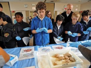
- A neuropathology station with real sheep brains and microscope slides of various tumor types.
- A rehabilitation and healing station where students learn what it’s like to apply and receive therapy.
- A suture station that gives students the chance to practice stitching.
- A brain and spine instrumentation station showing some of the hardware used in treatment.
- A research station where students can see and participate in DNA, tumor and laser experiments.
Students attending this year’s program come from diverse backgrounds and schools, including:
- The Dr. Betty Shabazz Delta Academy, a unique program for young women, designed to enhance or spark interest in math, science, technology and careers where minority women are scarcely represented.
- School on Wheels, Inc., which since 1993 has offered tutoring for homeless children.
- St. Francis of Assisi School, a Catholic elementary school with a student population composed mostly of Latino and Filipino students, with a small percentage of other ethnic groups.
- The James A. Foshay Learning Center, an Urban Education Partnership-affiliated Urban Learning Center, whose mission is to develop socially responsible citizens who are prepared to face the challenges of the 21st century.
“Brainworks,” which will be from 10 a.m. to 1:10 p.m. in the Harvey Morse Auditorium at Cedars-Sinai, includes lunch and is provided free by the Department of Neurosurgery and the Maxine Dunitz Neurosurgical Institute.