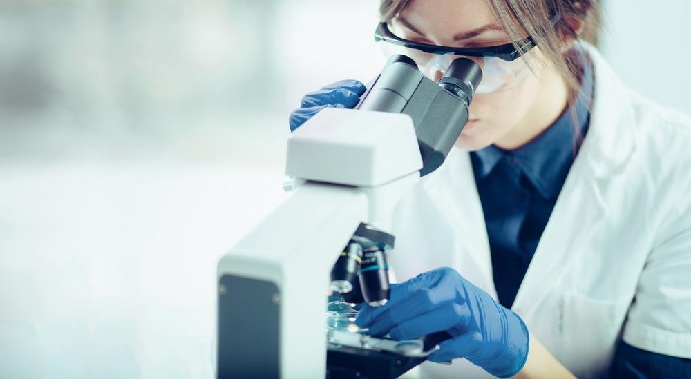
Microscopy techniques have revolutionized the way we perform medical diagnoses. Many diseases, such as cancers, cause changes at the cellular level that can be visualized using optical microscopy. For diagnosis of diseases such as malaria, that are caused by the presence of parasites, microscopes can be used to visualize the microscopic organisms.

Image Credit: Likoper/Shutterstock.com
A typical workflow involving microscopy for medical diagnosis involves taking some type of sample from the patient. This is usually in a form of a tissue biopsy or blood sample. Once the sample has been collected, it is labeled and sent for processing by a laboratory.
As diagnostic results are often required to inform clinical decision-making, diagnostic times ideally need to be as short as possible. Some biomarkers can undergo degradation during storage and transport, reducing the confidence in the final diagnosis.1 Delays in laboratory processing times can therefore increase waiting times for treatments, as well as having a negative impact on diagnosis quality and clinical decisions and outcomes.2
The value of diagnostic tests in improving treatment decisions and outcomes and the need to reduce sample processing times has led to a proliferation of research to develop point-of-care diagnostics.3 Some of these are in the form of new scanning technologies, but other developments focus on miniaturized analysis equipment for the evaluation of patient samples.
Point-of-Care Diagnostics
There are many potential advantages to point-of-care diagnostics. The most crucial one is the reduction in sample turnaround time to provide results for making informed clinical decisions. While it is unlikely the hospital laboratory will ever be replaced due to the diversity and increased sensitivity of many of the tests that can be performed there, more rapid screening could help alleviate the workloads of hospital laboratories, with additional testing only required for more complex or ambiguous cases.
One aspect of point-of-care diagnostic tests is that the results must be interpretable by clinicians and other non-laboratory staff and the test must be easy enough to perform that the staff do not need extensive additional training. One of the challenges with using microscopy for diagnosis is that the skill level and experience of the operator can often have a significant influence on the quality of the diagnosis.4
Compact Microscopy
There are several approaches to dealing with the challenge of image interpretation in microscopy. One is to use some type of automated image recognition to aid with diagnosis, the other is design tests that are more straightforward to interpret.
Recent work from the University of Illinois Urbana-Champaign has shown that it is possible to use an affordable, portable microscope for the detection of microRNAs.5 MicroRNAs are single-stranded non-coding RNAs and the presence of these in the blood is indicative of a variety of cancer types. Each cancer tends to have a unique microRNA associated with it.
The team could use its new device to diagnose prostate cancers successfully with imaging times ranging between 10 minutes to an hour. Detection limits for the device were 160 aM for particular nanoparticles on just a 30-minute array.
The microscope design was based on photonic resonator absorption microscopy. This is a label-free microscopy technique that exploits the strong interaction between nanoparticles and particular compounds such as RNA and then couples them to a photonic crystal to enhance the optical signals they produce in the experiment.
Gold nanoparticles interact with the microRNA. The resonant wavelength of the photonic crystal is then tuned to match the surface plasmons that form on the gold nanoparticle. This leads to localized quenching of the photonic crystal’s resonant reflection. What this means is that regions, where gold nanoparticles are present, show up as dark spots on the image.
Microscopy Methods
Photonic resonator absorption microscopy is an excellent technique for microRNA detection and requires no additional sample preparation, making it suitable for adaptation to a point-of-care method.
One of the main challenges that need to be overcome in the design of a photonic resonator absorption microscopy instrument is that large image areas are needed to compensate for the relative sparsity of the gold nanoparticles.
Larger image sizes mean the need for a greater focal area from the microscope as well as more complex image processing. However, it was possible to create a sufficient large sensor field of view, even with a webcam-quality CMOS sensor coupled with a low numerical aperture objective.
The enhancement of the optical signals from the gold nanoparticles meant signals could be recorded relatively efficiently, although the advantage of this label-free technique is that it is not subject to photobleaching of the fluorescent tags. This means it is possible to acquire long-exposure images.
Overall, the equipment could be assembled in less than a day for a total cost of $7000. The teams found that a red LED was sufficiently bright for sample illumination and offered the possibility for sufficiently short acquisition that diffusion kinetics and other effects could be studied.
Small, cost-effective devices with high selectivities, such as this microscope, are key developments in widening the range of tools we have for point-of-care measurements.
References and Further Reading
- Kristal, A. R., King, I. B., Albanes, D., Pollak, M. N., Stanzyk, F. Z., Santella, R. M., & Hoque, A. (2005) Short Communication Centralized Blood Processing for the Selenium and Vitamin E Cancer Prevention Trial : Effects of Delayed Processing on Insulin-Like Growth Factor Binding Protein 3 , Steroid Hormones , and Lymphocyte Viability. Cancer Epidemiology, Biomarkers & Prevention, 14, 727–731.
- Gill, D., Galvin, S., Ponsford, M., Bruce, D., Reicher, J., Preston, L., Bernard, S., Lafferty, J., Robertson, A., Rose-morris, A., Stoneham, S., Rieu, R., Pooley, S., Weetch, A., & Mccann, L. (2012) Laboratory sample turnaround times : do they cause delays in the ED ? Journal of Evaluation in Clinical Practice, 18, 121–127. https://doi.org/10.1111/j.1365-2753.2010.01545.x
- Yager, P., Domingo, G. J., & Gerdes, J. (2008) Point-of-Care Diagnostics for Global Health. Ann. Rev. Biomed. Eng, 10, 107–144. https://doi.org/10.1146/annurev.bioeng.10.061807.160524
- Crowley, R. S., Naus, G. J., III, J. S., & Friedman, C. P. (2003) Development of Visual Diagnostic Expertise in Pathology : An Information-processing Study. Journal of the American Medical Informations Association, 10(1), 39–51. https://pubmed.ncbi.nlm.nih.gov/12509356/
- Hosh, S. H. G., Antao, N. L. I., Iong, Y. A. X., Oung, Y. U., Athslag, M. I. P. R., Nal, E. G. E. G. O., Alkiewicz, E. R. F., Ohli, M. A. K., & Unningham, B. R. T. C. (2021) A compact photonic resonator absorption microscope for point of care digital resolution nucleic acid molecular diagnostics. Biomedical Optics Express, 12(8), 4637–4650.
Disclaimer: The views expressed here are those of the author expressed in their private capacity and do not necessarily represent the views of AZoM.com Limited T/A AZoNetwork the owner and operator of this website. This disclaimer forms part of the Terms and conditions of use of this website.