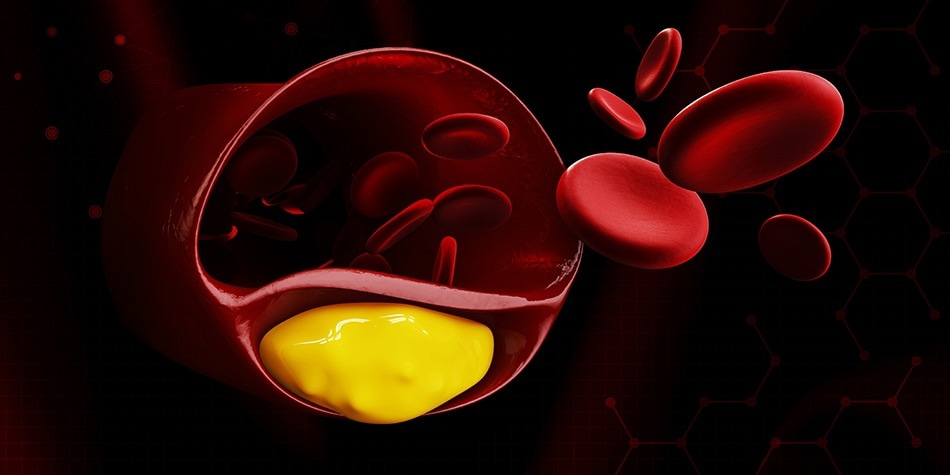
Coronary vascular diseases (CVDs) describe a group of disorders involving the heart and blood vessels, of which account for the most deaths for both men and women each year around the world1. One of the most common ways in which these diseases arise is due to the build-up of fatty deposits or plaque on the inner walls of the blood vessels; a disease otherwise known as atherosclerosis.
While conventional non-invasive diagnostic techniques exist, such as endovascular endoscopy, angioscopy, intravascular ultrasound, optical coherence tomography, and optical spectroscopy, in order to determine the degree of artherosclerotic diseases in coronary arteries exist; higher resolution options are in great demand2. While considered to be the most precise diagnostic tool available in the diagnosis of thrombogenic lesions and intravascular thrombi in coronary arteries, angioscopy techniques lack image quality and sensitivity.
In an effort to address this clinical need for an enhanced diagnostic tool in the determination of atherosclerotic lesions, researchers in the Department of Neurosurgery at the University of Michigan have developed a medical camera capable of providing a high resolution view of arteries. The research team, led by Dr. Luis Savastano M.D., developed a multimodal screening fibre endoscope (SFE) based on the original SFE design of Dr. Eric Seibel Ph.D. at University of Washington3, in which this tool has been used for early cancer detection purposes. While similar in design, the multimodal endoscope developed by Savastano’s team consist of a single optical fibre scanned by a piezoelectric drive that delivers red, blue and green laser beams to the tissue to collect the laser-induced reflectance and fluorescence in order to provide structural and biological analysis of vulnerable atherosclerotic arteries and veins2.
Mounted in an ultra-thin and highly flexible catheter, this endoscopic system consistently generates high-resolution images with a large field of focus potential that actively scans the entire arterial circumference. By simultaneously applying laser-induced fluorescence consisting of blue, green and red laser beams, whose measured wavelengths are 424, 488 and 642 nm, respectively, is scanned in a spiral pattern on the surface of the tissue. Once the laser beams are applied, subsequent laser reflectance and fluorescence is through a ring of optical fibres2. Due to the presence of intrinsic fluorochromes present in both healthy and diseased arteries and veins, these low power laser light sources are capable of collecting the reflected fluorescence, a method similar to that which is seen in conventional fluorescence spectrometry techniques.
Multimodal images obtained from the endoscope are then reconstructed and merged together in real time in order to generate the high-resolution videos used for diagnostic purposes. These videos are taken from the entire field of view of the endoscope at a resolution measured to be approximately 25 times greater than the standard endoscope, allowing for the generation of sharper images of vascular surfaces without the need to invasively examine vessels and risk possible plaque rupture within the arteries upon exposure2.
The original research performed by the team at Michigan Medicine found this technique to be especially powerful in the identification of surface defects in complicated plaques, even in cases where routine stroke work-up studies performed by the clinician had not elicited any concerns regarding the disease’s presence. Further, the researchers were able to classify their collected images into four categories of early, intermediate, advanced and complicated plaques, further supporting the potential diagnostic capability of this instrument for all stages of atherosclerotic disease. In fact, researchers found that the endo-luminal view provided by this method of angioscopy provided details regarding biologically unstable regions, with regards to the location of plaque in the arteries, therefore adding an additional value of this technique as a supplemental instrument in the local therapy of atherosclerosis in areas of higher vulnerability2.
While researchers suggest that carotid arteries, which are generally larger, straight vessels, represent the safest target to date for the initial in-human use of intravascular SFE, this forward-viewing endoscopic device is capable of generating impeccable images demonstrating the structural, chemical and biological status of vascular surfaces within the body.
References
- "Cardiovascular Diseases (CVDs)." World Health Organization. Sept. 2016.
- Luis E. Savastano, Quan Zhou, Arlene Smith, Karla Vega, Carlos Murga-Zamalloa, David Gordon, Jon McHugh, Lili Zhao, Michael M. Wang, Aditya Pandey, B. Gregory Thompson, Jie Xu, Jifeng Zhang, Y. Eugene Chen, Eric J. Seibel, Thomas D. Wang. Multimodal laser-based angioscopy for structural, chemical and biological imaging of atherosclerosis. Nature Biomedical Engineering, 2017; 1 (2): 0023. Web.
- Michigan Medicine - University of Michigan. "Laser-based camera improves view of the carotid artery." ScienceDaily. ScienceDaily, 10 February 2017. www.sciencedaily.com/releases/2017/02/170210130918.htm.
Disclaimer: The views expressed here are those of the author expressed in their private capacity and do not necessarily represent the views of AZoM.com Limited T/A AZoNetwork the owner and operator of this website. This disclaimer forms part of the Terms and conditions of use of this website.